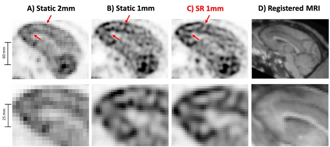
Researchers at CMITT have recently published an article on improving the resolution of brain positron emission tomography (PET) images using a technique called super-resolution (SR). SR aims to improve image quality by leveraging multiple acquisitions of the same target with known sub-resolution shifts. To achieve this, the researchers developed an SR estimation framework specifically for brain PET scans. They utilized a high-resolution infrared tracking camera to accurately measure and continuously track the shifts in real-time. By incorporating the tracking data into a PET reconstruction algorithm, they were able to correct motion and obtain PET images with visibly increased spatial resolution compared to standard static acquisitions. The study conducted experiments using moving phantoms and non-human primates, and quantitative analysis validated the effectiveness of the SR reconstruction method in improving visualization of small structures in brain PET scans. Enhanced PET resolution holds the potential for better detection of neurological disorders like Alzheimer’s disease, as well as providing a more accurate estimation of image-based input functions for quantifying dynamic brain PET studies.
Chemli Y, Tétrault MA, Marin T, Normandin MD, Bloch I, El Fakhri G, Ouyang J, Petibon Y. Super-resolution in brain positron emission tomography using a real-time motion capture system. Neuroimage 2023 272 ():120056