Outreach
Outreach involving training is at the foundation of our CMITT program. Click on the sections below to stay up to date on training opportunities.
Training in Medical Imaging
Training in Medical Imaging
CMITT provides research training in medical imaging, as currently applied to disciplines such as nuclear medicine (PET and SPECT), magnetic resonance imaging (MRI) and computed tomography (CT). The goal of the training is to provide an avenue for doctoral scientists in physics, engineering, mathematics, statistics, as well as related disciplines to enter and be successful in medical imaging research. This is achieved by providing training in radiological sciences to trainees with a strong quantitative background via structured didactic courses and seminars, enabling graduates to critically evaluate the field and formulate their own research ideas. Trainees participate in leading-edge research, with the opportunity to interact with a world-class faculty in a setting that combines the resources of the Harvard Medical School Joint Program in Nuclear Medicine (HMS-JPNM), the Harvard-affiliated Teaching Hospitals (e.g., Massachusetts General Hospital (MGH), Children’s Hospital Boston (CHB)), Brigham and Women’s Hospital, and the Harvard-MIT Division of Health Sciences and Technology (HST). CMITT stresses the methodology needed to advance medical imaging research specifically (in contrast to research training in imaging probes, animal models, or the study of disease mechanisms). The major foci of the didactic program are: (1) the physics of image formation with radiation, magnetic resonance and computed tomography, (2) the use of image processing to enhance the quantitative diagnostic and therapeutic capabilities of medical imaging, (3) the kinetic modeling of physiological processes needed to test hypotheses with cross-sectional PET, MRI and fMRI, as well as proton therapy and to advance quantitative functional imaging and molecular medicine.
Trainees can directly participate in unique training where trainees learn PET/MR theory, techniques, the underlying technical methods, and a wide range of clinical applications.
We will also provide educational and hands-on training to medical students, graduate students, and postdoctoral fellows and leverage the extended resources of our PET/MR program. In addition, the PET/MR program at MGH has attracted a large number of basic and clinical researchers, visiting scientists, and students from all around the world.
Fellows and Junior Faculty in training
- Kuang Gong, Ph.D.
- Paul Han, Ph.D.
- Nicolas Guehl, Ph.D.
- Rita Lahoud, M.D.
- Tao Sun, Ph.D.
- Thibault Marin, Ph.D.
- Maëva Dhaynaut, Ph.D.
- Amal Tiss, Ph.D.
- Yanis Chemli, MS
- Yanis Djebra, MS
Contact CMITT about training with us
Contact CMITT about training with us
Local Courses
Local Courses
A listing of local courses available through Harvard Medical School and the Harvard-MIT Health Sciences and Technology program can be found on the Gordon Center website:
Short Courses
Short Courses
CMITT will host an annual conference and workshop at the Society of Nuclear Medicine and Molecular Imaging for dissemination of results and new concepts. The program will include speakers from the Center with hands on sessions to introduce new technologies being developed. It will also include speakers from the imaging and industrial research communities that are focused on the technology at hand.
In addition to the annual conference, two to four short courses will be offered each year at MGH detailing the methods developed under CMITT.
Upcoming
P-41 Symposium at the World Molecular Imaging Congress (WMIC)
Prague, Czechia
September 5th – September 9th 2023
Sat, September 09
7:30 AM – 9:00 AM
Club E
The National Institute of Biomedical Imaging and Bioengineering (NIBIB) supports a large network of National Centers for Biomedical Imaging and Bioengineering (NCBIB) through the P41 grant mechanism. Three of these Centers comprise the faction of Molecular Imaging Technology Centers:
- The Center for Molecular Imaging Technology & Translation (CMITT), Massachusetts General Hospital, led by Dr. Georges El Fakhri, aims to develop and apply new imaging technologies that will revolutionize the way scientists and physicians view and use PET and MRI.
- The Resource for Molecular Imaging Agents in Precision Medicine, Johns Hopkins University, led by Dr. Martin Pomper, encompasses projects that extend from the development of reagents to detect and promote an immune reactive tumor microenvironment to the synthesis of nanodrones to treat cancer and combined small-molecule diagnostic and theranostic agents.
- The PET Radiotracer Translation and Resource Center (PET-RTRC), Washington University School of Medicine, led by Dr. Robert Gropler, seeks to develop new PET radiotracers that will image biologic targets modulating the ubiquitous disease processes of inflammation and oxidative stress.
The synergistic model of the P41 mechanism interaction of service and expertise within the Centers and other collaborating laboratories ensures other researchers may gain access to the newest Molecular Imaging Technology. During this session, participants will learn about cutting-edge molecular imaging innovations, in addition to the collaborating, service, and training opportunities disseminating from the three complementary, but non-overlapping programs that may benefit your own research and projects.
The 2022 annual workshop will be on deploying motion correction methodology in research and clinical environments. The workshop will include theoretical training on the PET/MR technology and hands-on training on the tools.
Short courses at IEEE Nuclear Science Symposium and Medical Imaging Conference
Full Day Course, IEEE Nuclear Science Symposium and Medical Imaging Conference, Boston, MA, USA, Nov. 2, 2020
PET/SPECT/MR short course
Coordinator/Course Instructor: Georges El Fakhri,
- SPECT&PET Basic Instrumentation & Image Formation (Georges El Fakhri, Ph.D., MGH, Boston)
- MR Basics (Chao Ma, PhD, MGH, Boston)
- PET/MR & SPECT/PET/CT Instrumentation (Hamid Sabet, MGH, Boston)
- Image Reconstruction and Quantitation (Joyita Dutta, MGH & UML, Boston)
- Clinical Applications of PET/MR (Georges El Fakhri, Ph.D., MGH, Boston)
Full Day Course, IEEE Nuclear Science Symposium and Medical Imaging Conference, Manchester, United Kingdom, 2019, Oct. 27 – Nov 3.
PET/SPECT/MR short course
Coordinator/Course Instructor: Georges El Fakhri,
Monday, 28 October 2019/ 8:00 AM – 5:00 PM
- Probes & Image Formation (Georges El Fakhri, Ph.D., MGH, Boston)
- Magnetic Resonance (Chao Ma, PhD, MGH, Boston)
- Instrumentation and system Design (Hamid Sabet, MGH, Boston)
- Image Reconstruction and Quantitation (Joyita Dutta, MGH & UML, Boston)
- Clinical Applications of PET/MR (Ruth Lim, M.D., MGH, Boston)
Short courses at the 2018 IEEE Nuclear Science Symposium and Medical Imaging Conference in Sydney, Australia, Nov 10-17
PET/MR: Principles and Applications
Sunday, 11 November 2018 / Time 8:00 am to 5:00 pm
- Principles of PET/MR and requirements for their integration (Georges El Fakhri, Ph.D., DABR)
- MR Basics (Chao Ma, PhD)
- Instrumentation and system Design for integration: small animal to human PET/MR (Armin Kolb, PhD)
- Challenges and benefits of integration I: Attenuation correction, Motion correction (Quanzheng Li, Ph.D.)
- Challenges and benefits of integration II: Dual Probe PET/MR & Imaging of multiple physiological processes (Georges El Fakhri, Ph.D., DABR)
- Clinical Applications of PET/MR (Ruth Lim, M.D.)
Short courses at the Radiological Society of North America (RSNA)
Short Courses, 2020 RSNA Annual Meeting, Chicago, IL.
Applications of Deep Learning in MRI and PET/MRI Image Formation
Fang Liu, PhD, Boston, MA
Learning Objectives
1) Present a technical overview of DL in medical imaging and discuss some recent DL applications that successfully translate new
learning-based approaches into performance improvement in MR and PET/MR imaging workflow. 2) Draw tightly connections
between fundamental DL concepts and technical challenges in medical imaging. 3) Cover rapid image acquisition and reconstruction
to image post-processing such as image segmentation and synthesis in MR and PET/MR. 4) Discuss open problems in DL that are
particularly relevant to medical imaging and the potential challenges and opportunities in this emerging field.
Physics & Technology
Georges El Fakhri, PhD, Boston, MA
Learning Objectives
1) Understand current technology both in terms of instrumentation and imaging science as they pertain to PET/MR. 2) Understand
the potential applications afforded by PET/MR technology developments (e.g., instrumentation, AI).
Short Courses, 2019 RSNA Annual Meeting, Chicago, IL.
Emerging Technology: PET/MRI Update 2019
Organizer/Moderator: Rathan Subramanian, Georges El Fakhri
Thursday December 5th 2019 / 16:00 – 18:00, Course # RC717
- 16:00 – 16:20 PET/MRI 2019: Clinical Practice Implementation
- Geoffrey B. Johnson, MD, PhD, Rochester MN
- 16:20 – 16:40 PET/MRI Clinical Applications: Brain and Head and Neck
- Alexander Drzezga, MD, Cologne, Germany
- 16:40 – 17:00 PET/MRI Clinical Applications: Body
- Thomas A. Hope, MD, San Fransisco, CA
- 17:00 – 17:20 PET/MRI Clinical Applications: Cardiac
- Pamela K. Woodard, MD, Saint Louis, MO
- 17:20 – 17:40 PET/MRI Clinical Applications: Pediatrics
- Lisa States, MD, Philadelphia, PA
- 17:40 – 18:00 PET/MRI 2019: Physics and Instrumentation
- Georges El Fakhri, PhD, Boston, MA
Short Courses, 2018 RSNA Annual Meeting, Chicago, IL
Emerging Technologies: PET/MRI update 2018
Organizer/Moderator: Rathan Subramanian, Georges El Fakhri
Wednesday Nov. 28, 8:30-10:00 AM | RC517 | Room: S505AB
- 14:00 –14:10 PET/MRI: The Evolving Imaging Field of Structure & Function
Rathan M. Subramaniam, MD, PhD - 14:10 –14:40 PET/MRI Physics: The Opportunities and Challenges
Georges El Fakhri, PhD - 14:40 –14:55 PET/MRI Clinical Applications: Brain and Head and Neck
Alexander Drzezga, MD - 14:55 –15:10 PET/MRI Clinical Applications: Body
Thomas A. Hope, MD - 15:10 –15:30 PET/MRI Clinical Applications: Cardiac
Pamela K Woodard, MD
Categorical Courses at the Society of Nuclear Molecular Imaging (SNMMI) Meetings
SNMMI 2021 Mid-Winter Meeting, Virtual
Artificial Intelligence in Nuclear Medicine, Sponsored by the SNMMI Physics
Instrumentation and Data Sciences Council
Organizer: Joyita Dutta
2:15–3:45 pm ET
Session Description
This session will focus on the fundamentals of artificial intelligence (AI) and highlight recent advances in deep learning in the context of nuclear medicine imaging. Deep learning is particularly well-suited for the analysis of large and complex datasets including images. Unlike classical machine learning approaches, which rely on predetermined handcrafted features, deep learning algorithms learn features that are most meaningful for a given task. In this session, we aim to provide both a foundational overview of AI methodology and discuss major applications of AI in nuclear medicine.
The session is intended to provide the attendees with an in-depth understanding of AI tools that have the potential to significantly improve diagnosis, prognosis, and treatment response assessment, and to take a significant step toward realization of precision (personalized) medicine.
Session Objectives
- Describe the framework of a deep learning algorithm
and how this differs from classical machine learning. - Examine motivations for applying machine learning
and deep learning methods to radiological images,
and how these may significantly improve diagnosis,
prognosis, and treatment response assessment. - Recognize the pitfalls and challenges of, as well as
solutions towards, the translation of deep learning
methods to routine clinical practice
SNMMI 2020 Annual Meeting – Virtual
Simultaneous PET/MRI: Advantages and Clinical Applications
Georges N. El Fakhri, PhD, FSNMMI; Hongyu An, PhD; Kuang, Gong, PhD; Helen R. Nadel, MD FRCPC
Tuesday Sept. 8, 2020 1:00 PM – 2:00 PM
Course Learning Objectives
Compare and better understand the recent developments in instrumentation and image analysis in PET/MRI
Better understand the upcoming role of machine learning in PET/MRI.
Examine the role of PET/MRI in the context of oncologic, brain and pediatric PET/MRI
Categorical Courses, 2019 SNMMI Annual Meeting, Anaheim, CA.
Advances in Simultaneous PET/MRI Imaging and Clinical Applications
Organizer/Moderator: Georges El Fakhri, Richard Laforest
Sunday June 23, 2019 / 16:30 – 18:30
- 16:30 – 17:00 PET/MRI: Technology
- Richard Laforest, PhD
- 17:00 – 17:30 Application of Machine Learning in PET/MRI
- Hongyu An, PhD
- 17:30 – 18:00 Pediatric PET/MRI
- Lisa J. States, MD
- 18:00 – 18:30 Cardiovasular PET/MR imaging
- Georges El Fakhri, PhD, FIEEE, FAAPM, FSNMMI
AI for Data Sciences
Organizer/Moderator: Georges El Fakhri
Tuesday June 25, 2019 / 15:00 – 16:30
- 15:00 – 15:30 AI in PET and MR
- Georges El Fakhri, PhD, MGH, Boston
- 15:30 – 16:00 Deep Learning in Tomographic Reconstruction
- Quanzheng Li, PhD, MGH, Boston
- 16:30 – 9:00 Promise and Challenes of Data Science
- Mark Michalski, MD, MGH, Boston
Categorical Courses, 2018 SNMMI Annual Meeting, Philadelphia, PA.
Advances in Simultaneous PET/MRI Imaging and Clinical Applications
Organizer/Moderator: Georges El Fakhri, Richard Laforest
Sunday June 22, 2018 / 16:30 –18:30 EDT
- 4:30 PM -5:00 PM PET/MRI: Technology
Richard Laforest, PhD - 5:00 PM -5:30 PM Application of Machine Learning in PET/MRI
Hongyu An, PhD - 5:30 PM –6:00 PM Pediatric PET/MRI
Lisa J. States, MD - 6:00 PM -6:30 PM Cardiovasular PET/MR imaging
Georges El Fakhri, PhD, FIEEE, FAAPM, FSNMMI
PET/CT and PET/MR for Adults and Children: Fundamental and Evolving Practices. Instrumentation Update: PET/MR
Saturday June 23, 2018 / 7:30 AM- 10:00 PM EDT
- 8:00 AM – 8:20 AM Machine Learning and PET: A Great Combination?
Vincent Gaudet, PhD, PEng, SMIEEE - 8:20 AM – 8:40 AM Instrumentation Update: New Technologies for PET/CT
Suleman Surti, PhD - 8:40 AM – 9:00 AM Instrumentation Update: PET/MR
Georges N. El Fakhri, PhD - 9:00 AM – 9:20 AM A Guide to Dosimetry: PET/CT vs. PET/MR
Richard Laforest, PhD - 9:20 AM – 9:40 AM Image Gently Update
Frederic H. Fahey, DSc, FSNMMI
ISMRM 2020
Session Topic: Gliomas, MR/PET/MRS for Guiding Radiotherapy
Georges El Fakhri
Saturday, 8 August 2020, 15:00 – 15:30 UTC
– Demonstrate uses of MR/PET/MRS in guiding the delivery of raidation therapy
Future workshops, seminars, short courses
- November 2022: PET/MRI workshop at virtual IEEE NSS/MIC
- TBD: PET/MR Workshop at Massachusetts General Hospital
- Recent advances in kinetic modeling of receptor/neurotransmitter systems in non-steady state
- Deployment of joint estimation and reconstruction for simultaneous PET and MR clinical and preclinical data are another dimension for methodological development
Visiting Scientist Program
Visiting Scientist Program
Visiting Scientist Program
A small number of participants will be provided in-depth training in specific PET/MR technology.
If you are interested in participating in this program, please fill out the contact form below:
Electronic Teaching Materials
Electronic Teaching Materials
Videotaped lectures are part of the CMITT effort, along with the Gordon Center for Medical Imaging, to disseminate the latest research topics in medical imaging as it pertains to simultaneous PET/MR. Additional lectures can be found here.
- Medical Imaging Sciences and Applications HST Course
- This course provides a general overview of Medical Imaging, and the webpage includes video lectures on many of the topics
- Basics of MR
- Basics of PET
- Basics of Kinetic Modeling
- PET/MR: Challenges and Opportunities
- Coming soon: Joint PET/MR reconstruction
- Coming soon: How to use MR for motion correction
Software Resources
Software Resources
MRiLab A Numerical Magnetic Resonance Imaging (MRI) Simulation Platform
Code: https://github.com/leoliuf/MRiLab
The MRiLab is a numerical MRI simulation package. It has been developed and optimized to simulate MR signal formation, k-space acquisition and MR image reconstruction. MRiLab provides several dedicated toolboxes to analyze RF pulse, design MR sequence, configure multiple transmitting and receiving coils, investigate magnetic field related properties and evaluate real-time imaging technique. The main MRiLab simulation platform combined with those toolboxes can be applied to customize various virtual MR experiments which can serve as a prior stage for prototyping and testing new MR technique and application.
Questions? Contact Fang Liu
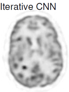
IterativeCNN: Iterative PET image reconstruction using convolutional neural network representation
Code: https://github.com/zgongkuang/IterativeCNN
This code and shared simulation datasets can be used to reproduce the simulation results published in our work, and also help the community further develop approaches combining deep learning and iterative reconstruction. Below is the abstract of this work.
PET image reconstruction is challenging due to the ill-poseness of the inverse problem and limited number of detected photons. Recently, the deep neural networks have been widely and successfully used in computer vision tasks and attracted growing interests in medical imaging. In this work, we trained a deep residual convolutional neural network to improve PET image quality by using the existing inter-patient information. An innovative feature of the proposed method is that we embed the neural network in the iterative reconstruction framework for image representation, rather than using it as a post-processing tool. We formulated the objective function as a constrained optimization problem and solved it using the alternating direction method of multipliers algorithm. Quantification results show that our proposed iterative neural network method can outperform the neural network denoising and conventional penalized maximum likelihood methods.
Questions? Contact Kuang Gong
Matlab processing for pharmacokinetic PET studies in NHP and Humans
We have developed an analysis pipeline in Matlab for processing pharmacokinetic PET studies performed in non-human primates and in humans. The workflow includes registration of the images to a standardized template space, automated extraction of regional brain time activity curves (TACs) using a set of brain atlases, processing of blood and plasma data, if available, application of kinetic models for accurate quantification of the physiological processes of interest and computation of parametric maps.
The list of implemented mathematical models includes blood-based techniques such as full compartment models in different configurations (one-tissue or two-tissue in reversible and irreversible modes with and without inclusion of the vascular contribution to the PET signal as a model parameter), graphical methods (such as Logan, MA1 and Patlak) and reference-tissue based techniques (such as SRTM, Logan DVR, MRTM). The inputs that are required for the pipeline consists of the individual structural MR images and the dynamic PET images either in dicom or in nifti format. If blood-based models are selected, the user also needs to provide the whole-blood and plasma concentrations measured over the course of the study as well as corresponding parent fraction measurements for generation of metabolite-corrected arterial input function.
Questions? Contact Nicolas Guehl
LTSA-MRI
A manifold learning-based method that leverages the intrinsic low-dimensional structure of MR Spectroscopic Imaging (MRSI) signals for joint spectral quantification.
The available code consists of MATLAB®-based scripts and functions along with corresponding example simulated datasets.
Please email developer Chao Ma for help and comments.
C. Ma, et. al. “Joint spectral quantification of MR spectroscopic imaging using linear tangent space alignment (LTSA)-based manifold learning” Magn Reson Med. 2022 Nov 20;. doi: 10.1002/mrm.29526.
PLEASE NOTE: The software available on this page is provided free of charge and comes without any warranty. CMITT and the MGH/HMS do not take any liability for problems or damage of any kind resulting from the use of the files provided. Operation of the software is solely at the user’s own risk. The software developments provided are not medical products and must not be used for making diagnostic decisions.
The software is provided for non-commercial,, non-clinical academic use only. Usage or distribution of the software for commercial purpose is prohibited. All rights belong to the authors and/or the Massachusetts General Hospital. If you use the software for academic work, please give credit to the author in publications and cite the related publications.
Some of the software developments provided are still work-in-progress such that the software is not completely free of errors.
Gordon Lecture Series
Gordon Lecture Series
Regular seminars are held on Fridays with experts from around the world presenting their research
Invited Talks
Invited Talks
2017: Invited speaker; SNMMI Mid Winter Meeting – Phoenix, AZ,
“Role of Quantification in Amyloid and Tau Imaging”.
2017: Invited speaker; SNMMI Mid Winter Meeting – Phoenix, AZ,
“Role of PET/MR in Cancer and Non-Oncologic Application”.
2017: Invited Grand Round Speaker: University of Chicago, Chicago, IL:
“Quantitative PET/MR in oncologic, cardiac and brain applications”.
2017: Invited Keynote Speaker: Quebec Bio-Imaging Network, MNI, Montreal, CAN:
“Simultaneous PET/MR Lessons learned”.
2017: Invited speaker. University of Dartmouth PET-CT Symposium:
“PET/MR: Strengths & Challenges”. Stowe, VT.
2017: Invited speaker. University of Dartmouth PET-CT Symposium:
“Dose Reduction in PET studies”. Stowe, VT.
2017: Invited speaker. Society of Pediatric Radiology (SPR) annual Meeting, Vancouver, CN:
“Physics & Instrumentation: Considerations for Pediatric PET/MR”.
2017: Invited speaker: Ministry of Health, Kuwait:
- “Basic atomic and nuclear physics”
- “Decay of radioactivity”
- “Scintillation Detectors: Non-imaging”
- “Gamma camera: Basics, operations, consideration”
- “SPECT , filtering, processing”
- “Hybrid imaging: Basic CT”
- “Basic radiation protection in nuclear medicine”
2017: Invited Speaker: International Society of Magnetic Resonnance in Medicine (ISMRM), Honolulu, HI:
“Advanced Multimodality Cardiac Imaging: PET/MR”
2017: Invited Speaker: Stanford University, Palo Alto, CA:
“PET/MR for in vivo mapping of cancer, brain and heart function.”
2017: Plenary Speaker: Frontiers in Medical Imaging Symposium, Jiao Tong University, Shanghai (2 Plenary speakers: NIBIB Director Dr Pettigrew and Dr El Fakhri).
2017: Invited speaker; SNMMI Annual Meeting, Denver, CO:
“Basic Science Plenary Session: Data Analysis & Instrumentation”.
2017: Invited lecture: SNMMI Annual Meeting, Denver, CO:
“Decreasing PET Radiation Dose Using Currently Available Technology”.
2017: Invited Speaker: Tsinghua University, Beijing:
PET/MR for in vivo mapping of cancer, brain and heart function.
2017: Invited Plenary Speaker, Fully 3D Meeting, Xi’ian: Quantitative PET/MR for in vivo mapping of cancer, brain and heart function.
2017: Invited speaker: Yonsei University, Seoul, Korea:
Simultaneous PET/MR: Challenges and Opportunities for cardiac, oncologic and neurologic imaging.
2017: Invited speaker: KAIST University, Seoul, Korea:
Simultaneous PET/MR: Challenges and Opportunities for Engineers.
2017: Invited Keynote speaker: Molecular Imaging Instrumentation Workshop, The University of Chicago in Hong Kong:
“PET/MR in medicine and biology”.
2017: Invited speaker: National Academy of Science, Taipei, Taiwan:
“PET/CT and PET/MR in radiation therapy”.
2017: Invited speaker: National Academy of Science, Taipei, Taiwan:
“PET/CT and PET/MR in radiation therapy”.
2017: Invited Keynote speaker: French Naional Nuclear Medicine Society (APRAMEN), Paris, France:
“Quantitative PET/MR for in vivo functional Cardiac, Neurologic and Oncologic Imaging”.
2017: Invited Speaker: Radiological Society of North America (RSNA) Annual Meeting, Chicago IL:
“PET/MR Physics and Instrumentation: Opportunity and Challenges”
2017: Invited Keynote speaker: Bangladesh Atomic Energy Commission, Medical Sciences, Dhaka, Bangladesh.
“Recent Developments in PET-CT & PET-MR: Technical and Clinical Considerations”.
2017: Invited Keynote Speaker, Memorial Sloan Kettering Cancer Center, New York, NY.
“Quantitative Oncologic and cardiac PET/MR: the MGH experience”.
2018: Invited Keynote Speaker, National Pakistani Medical Physics Meeting, Karachi, Pakistan:
“Recent Developments in PET/CT and PET/MR: Lessons learned”
2018: Invited Keynote Speaker, Kuwait Foundation for Advancement of Science, Nuclear Medicine Workshop, Kuwait:
“Quantitation of Myocardial Blood Flow with PET”
2018: Invited Keynote speaker: Molecular Imaging Instrumentation Workshop, The University of Chicago in Hong Kong:
“Simultaneous PET/MR in Cancer: lessons learned from sarcoma studies”.
2018: Invited speaker: National Taiwan University, Taipei, Taiwan:
“Cardiac PET/MR: Beyond Flow”.
2018: Visiting Professor Lecturship, Children’s Hospital of Philadelphia, Deaprtment of Radiology, Philadelphia, PA:
“Simulatneous PET/MR: Advances and Challenges”
2018: Invited Plenary Speaker: 2018 Asia Pacific Conference on Plasma and Terahertz Science, Xi’ian, China: “Exploring Pathophysiology using quantitative PET/MR”.
2018: Invited speaker; SNMMI Annual Meeting, Philadelphia, PA: Basic Science Plenary Session:
“Data Analysis & Instrumentation”.
2018: Invited Plenary Speaker: Royal North Sore Hospital, Sydney, Australia:
“Exploring Pathophysiology using quantitative oncologic, cardiac and brain PET/MR”.
2018: Invited Speaker: Radiological Society of North America (RSNA) Annual Meeting, Chicago IL:
“PET/MR Physics and Instrumentation: Opportunity and Challenges”
2018: Invited Plenary Speaker: BOE Innovator Lecture Series, Beijing, China:
“Role and Opportunities for Molecular Imaging & AI in Healthcare”
2018: Invited Plenary Speaker: University of British Columbia, Vancouver, Canada:
“Imaging of Oncologic, Cardiac, and Brain Patho-Physiology Using Positron Emission Tomography and Magnetic Resonance”
2019: Invited Speaker: US Food and Drug Administration (FDA), Silver Spring, MD:
“PET/MR and New Technologies”
2019: Invited Grand Rounds Speaker: University of British Columbia, Canada:
“Imaging of oncologic patho-physiology using PET and MR”.
2019: Invited Grand Rounds Speaker: Tiantan University, Beijing, China:
“Molecular Imaging of brain in normal function and disease using PET and MR”
2019: Invited Plenary Speaker: Zhengzhou University, Department of Pharmaceutical Sciences, China:
“Radiochemistry for PET/MR imaging of cardiac and cancer disease”.
2019: Invited Plenary Speaker: National Institutes of Health, Taipei, Taiwan:
“Probing Patho-physiology with PET/MR”.
2019: Invited Plenary Speaker: Intercontinental Nuclear Insttitute Meeting, Boston, MA:
“Application of Nuclear Technology to Medical Imaging at the Gordon Center for Medical Imaging”.
2019: Invited Plenary Speaker: Chemistry Department, Zhengzhou University, China:
“Quantitative Molecular Simultaneous PET/MR: Are we there yet?”.
2019: Invited Plenary Speaker: Henan Hospital, Zhengzhou, China:
“Molecular PET/MR: Present and Future”.
2019: Invited Plenary Opening Lecture Speaker: Molecular Imaging Instrumentation Workshop, The University of Chicago in Hong Kong, China:
“Quantitative PET/MR: New frontier for molecular imaging”.
2019: Invited Speaker, Annual Meeting of the American Association of Phycisists in Medicine (AAPM), San Diego, TX.
“Dose optimization in PET/CT and PET/MR”.
2019: Invited Speaker, Annual Meeting of the American Association of Phycisists in Medicine (AAPM), San Diego, TX.
“Reducing sedation and anesthesia in PET/CT and PET/MR”.
2019: Invited Keynote Plenary Speaker: Annual Meeting of the Asian Society of Magnetic Resonnance in Medicine (ASMRM), Shanghai, China:
“Brain Molecular Imaging of Normal Function and Disease”.
2019: Invited Keynote Plenary Speaker: Annual Meeting of the Chinese Society of Magnetic Resonnance in Medicine (CSMRM), Shanghai, China:
“Molecular Imaging in Cardiac & Oncologic Applications”.
2019: Invited Speaker: International Society of Magnetic Resonnance in Medicine (ISMRM), Montreal, Canada:
“CNS tumors: Molecular Imaging with PET/MR”.
2019: Invited Speaker: International Society of Magnetic Resonnance in Medicine (ISMRM), Montreal, Canada:
“Writing / Getting great research grants”.
2019: Edward J. Hoffman Lectureship, Societyof Nuclear Medicine and Molecular Imaging (SNMMI), Anaheim, CA:
“Probing patho-physiology with molecular imaging”
2019: Invited Plenary speaker; SNMMI Annual Meeting, Anaheim, CA: Basic Science Plenary Session:
“Data Analysis & Instrumentation”.
2019: Invited speaker, Society of Nuclear Medicine and Molecular Imaging (SNMMI), Anaheim, CA:
“AI in Medical Imaging”
2019: Invited Plenary Keynote Speaker: International Society of Magnetic Resonnance in Medicine (ISMRM) Workshop, Zhengzhou, China:
“Brain Molecular Imaging: From Normal Function to Disease State”.
2019: Invited Plenary Speaker: Nanjing General Hospital, Nanjing, China:
“Oncologic Molecular Imaging Using PET/MR”.
2019: Invited Speaker: 2019 Ultra High Sensitivity PET, Linyi, China:
“Brain Molecular Imaging”.
2019: Invited Plenary Speaker: International Biomedical Engineering Symposium (IBES), Incheon, Korea: “Innovation & Translation in Molecular Imaging in the era of Big Data”.
2019: Invited Plenary Speaker: 2nd International Conference on Medical Imaging, Boston, MA:
“Imaging Patho-Physiology with PET and MR”.
2019: Invited Speaker: Radiological Society of North America (RSNA) Annual Meeting, Chicago IL:
“PET/MR Physics”.
2020: Invited Plenary Speaker: Tsinghua Sanya International Mathematics Forum, Sanya, China:
“Molecular Imaging in the era of AI”.
2020: Invited Speaker: Society of Nuclear Medicine and Molecular Imaging, Mid-Winter Meeting, Tampa, FL:
“Brain PET in the era of AI”.
2020: Invited Speaker: University College London, London, UK:
“Physiological PET/MR: Beyond Reconstruction”.
2020: Invited Keynote Speaker: SPIE Medical Imaging, Houston, TX:
“Innovations and Translation in Molecular PET/MR and PET/CT”.
2020: Invited Grand Round Speaker: University of Chicago, Chicago, IL:
“Cardiac, Brain and Oncologic PET/MR”.
2020: Invited Speaker: Society of Nuclear Medicine and Molecular Imaging (SNMMI), Virtual Annual Meeting:
“PET/MR Instrumentation”.
2020: Invited Plenary speaker; Society of Nuclear Medicine and Molecular Imaging (SNMMI), Virtual Annual Meeting:
“Basic Science Plenary Session: Data Analysis & Instrumentation”.
2020: Invited Speaker: International Society of Magnetic Resonnance in Medicine (ISMRM), Virtual:
“PET/MR/MRSI for Image Guided Radiation Therapy”.
2020: Invited Speaker: American Society of Nuclear Cardiology (ASNIC) Annual Meeting, Virtual: “Advanced Strategies for PET Cardiac Quantitation”.
2020: Invited Speaker: Society of Pediatric Radiology (SPR) Annual Meeting, Virtual:
“AI in PET/MR”.
2020: Invited Speaker: Radiological Society of North America (RSNA) Annual Meeting, Chicago IL (Virtual):
“PET/MR Physics and Instrumentation”.
2020: “Deep learning for image segmentation: magnetic resonance imaging” IEEE Nuclear Science Symposium & Medical Imaging Conference
2020: “Applications of deep learning in MRI and PET/MRI image formation” RSNA Scientific Assembly & Annual Meeting.
2020: Invited speaker: “Machine (Deep) Learning for MSK: image acquisition & reconstruction” ISMRM & SMRT Virtual Conference & Exhibition
2021: Invited Plenary speaker; Society of Nuclear Medicine and Molecular Imaging (SNMMI), Virtual Annual Meeting: Basic Science Plenary Session: Data Analysis & Instrumentation”.
2021: Invited Keynote Speaker, New England American Association of Physicists in Medicine, Annual Meeting: “Molecular Imaging and AI in Radiation Therapy: Quo Vadis ?” (Virtual).
2021: Invited Speaker, 7th NIH Brain Initiative Meeting, “Molecular Imaging Brain PET and PET/MR” (Virtual).
2021: Invited Keynote Speaker, Emirates International Conference in Nuclear Medicine and Molecular Imaging. “PET/MR Imaging for guiding therapy” (Virtual).
2021: Invited Speaker: Radiological Society of North America (RSNA) Annual Meeting, Chicago IL (Hybrid): “PET/MR Physics and Instrumentation”.
2021: Invited Speaker: 4th Annual Cardiovascular Institute Symposium, Stanford University, CA (Hybrid): “Imaging membrane potential in vivo: First in Human studies”.
2022: Invited speaker: Ministry of Health, Kuwait: “Basic atomic and nuclear physics”
“Decay of radioactivity”
“Scintillation Detectors: Non-imaging”
“Gamma camera: Basics, operations, consideration”
“SPECT , filtering, processing”
“PET: Basics, attenuation correction, artifacts, etc”
“Hybrid imaging: Basic CT”
“Challenges and Opportunities afforded by PET/MR”
2021: Invited Speaker, Martinos Center for Biomedical Imaging, “Fast high-resolution metabolic imaging with SPICE”
2022: Invited Grand Round Speaker, UCLA Cardiovascular Distinguished Seminar Series: “Quantitative Mapping of Cardiac Membrane Potential”, Los Angeles, CA.
2022: Invited Keynote Plenary Speaker, Fast Timing in Mecical Imaging Conference, “Clinical and Technical Impact of Fast TOF PET”, Valencia, Spain.
2022: Invited Keynote Plenary Speaker, 4th Jagiellonian Symposium on Fundamental and Applied Subatomic Physics, “Clinical and Technical Considerations for Fast TOF PET”, Krakow, Poland.
2022: Invited Speaker, Mediterranean Thematic Workshop in Advanced Molecular Imaging, “Clinical and Technical Considerations for Fast TOF and for High Resolution PET”,Portorož, Slovenia.
Publication List
Publication List
Selected publications since establishment of CMITT
| Title | Authors | Journal | Date | Categories |
|---|---|---|---|---|
| Arterial spin labeled perfusion imaging with balanced steady-state free precession readout and radial sampling | Han PK, Marin T, Zhuo Y, Ouyang J, El Fakhri G, Ma | Magn Reson Imaging 2023 102 ():126-132 | 2023 | TR&D 1 |
| B inhomogeneity-corrected T mapping and quantitative magnetization transfer imaging via simultaneously estimating Bloch-Siegert shift and magnetization transfer effects | Jang A, Han PK, Ma C, El Fakhri G, Wang N, Samsonov A, Liu F | Magn Reson Med 2023 (): | 2023 | TR&D 1 |
| Impact of motion correction on [F]-MK6240 tau PET imagin | Tiss A, Marin T, Chemli Y, Spangler-Bickell M, Gong K, Lois C, Petibon Y, Landes V, Grogg K, Normandin M, Becker A, Thibault E, Johnson K, El Fakhri G, Ouyang J | Phys Med Biol 2023 68 (10): | 2023 | TR&D 1 |
| Super-resolution in brain positron emission tomography using a real-time motion capture system | Chemli Y, Tétrault MA, Marin T, Normandin MD, Bloch I, El Fakhri G, Ouyang J, Petibon Y | Neuroimage 2023 272 ():120056 | 2023 | TR&D 1 |
| Measurement of Cerebral Perfusion Indices from the Early Phase of [F]MK6240 Dynamic Tau PET Imaging | Guehl NJ, Dhaynaut M, Hanseeuw BJ, Moon SH, Lois C, Thibault E, Fu JF, Price JC, Johnson KA, El Fakhri G, Normandin MD | J Nucl Med 2023 64 (6):968-975 | 2023 | TR&D 3 |
| Human biodistribution and radiation dosimetry of the demyelination tracer [18F]3F4AP | Brugarolas P*, Wilks MQ*, Noel J, Kaiser JA, Vesper DR, Ramos-Torres KM, Guehl NJ, Macdonald-Soccorso MT, Sun Y, Rice PA, Yokell DL, Lim R, Normandin MD, El Fakhri G | Eur J Nuc Med Molec Imag. 2023; 50(2):344-351 | 2023 | TR&D 3 |
| PET imaging studies to investigate functional expression of mGluR2 using [11C]mG2P001 | Yuan G*, Dhaynaut M*, Guehl NJ, Neelamegam R, Moon S-H, Qu X, Poutiainen P, Afshar S, El Fakhri G, Brownell AL, Normandin MD | J Cereb Blood Flow Metab. 2023 Feb;43(2):296-308 | 2023 | TR&D 3 |
| Memory consistent unsupervised off-the-shelf model adaptation for source-relaxed medical image segmentation | Liu X, Xing F, El Fakhri G, Woo J | Med Image Anal. 2023 Jan;83:102641 | 2023 | TR&D 2 |
| Posterior estimation using deep learning: a simulation study of compartmental modeling in dynamic positron emission tomography | Liu X, Marin T, Amal T, Woo J, El Fakhri G, Ouyang J | Med Phys. 2023 Mar;50(3):1539-1548. | 2023 | TR&D 2 |
| Network Tau spreading is vulnerable to the expression gradients of APOE and glutamatergic-related genes | Montal V, Diez I, Kim CM, Orwig W, Bueichekú E, Gutiérrez-Zúñiga R, Bejanin A, Pegueroles J, Dols-Icardo O, Vannini P, El-Fakhri G, Johnson KA, Sperling RA, Fortea J, Sepulcre J | 2022 Jul 27;14(655):eabn7273 | 2022 | TR&D 3 |
| Kinetic evaluation and assessment of longitudinal changes in reference region and extracerebral [18F]MK-6240 PET uptake | Fu JF, Lois C, Sanchez J, Becker JA, Rubinstein ZB, Thibault E, Salvatore AN, Sari H, Farrell ME, Guehl NJ, Normandin MD, El Fakhri G, Johnson KA, Price JC | J Cereb Blood Flow Metab. 2022; 271678X221142139. | 2022 | TR&D 3 |
| Radiochemical synthesis and evaluation of 3-[11C]methyl-4-amino-pyridine in rodents and non-human primates for imaging potassium channels in the CNS | Sun Y, Guehl NJ, Zhou Y-P, Takahashi K, Belov V, Dhaynaut M, Moon S-H, El Fakhri G, Normandin MD, Brugarolas P | ACS Chem Neurosci. 2022;13(23):3342-3351 | 2022 | TR&D 3 |
| ACT: Semi-supervised Domain-adaptive Medical Image Segmentation with Asymmetric Co-Training | Liu X, Xing F, Shusharina N, Lim R, Kuo CJ, El Fakhri G, Woo J | Med Image Comput Comput Assist Interv. 2022 Sep;13435:66-76 | 2022 | TR&D 2 |
| Unsupervised Black-Box Model Domain Adaptation for Brain Tumor Segmentation | Liu X, Yoo C, Xing F, Kuo CJ, El Fakhri G, Kang JW, Woo J | Front Neurosci. 2022;16:837646 | 2022 | TR&D 2 |
| Manifold Learning via Linear Tangent Space Alignment (LTSA) for Accelerated Dynamic MRI with Sparse Sampling | Djebra Y, Marin T, Han PK, Bloch I, El Fakhri G, Ma C | IEEE Trans Med Imaging. 2022 Sep 19;PP. | 2022 | TR&D 1 |
| Joint spectral quantification of MR spectroscopic imaging using linear tangent space alignment-based manifold learning | Ma C, Han PK, Zhuo Y, Djebra Y, Marin T, El Fakhri G | Magn Reson Med 2022 | 2022 | TR&D 1 |
| Design, Synthesis, and Characterization of [18F]mG2P026 as a High-Contrast PET Imaging Ligand for Metabotropic Glutamate Receptor 2. | Yuan G, Dhaynaut M, Guehl NJ, Afshar S, Huynh D, Moon SH, Iyengar SM, Jain MK, Pickett JE, Kang HJ, Ondrechen MJ, El Fakhri G, Normandin MD, Brownell AL. | J Med Chem. 2022 Jul 28;65(14):9939-9954. | 2022 | TR&D 3 |
| Feasibility study of clinical target volume definition for soft-tissue sarcoma using muscle fiber orientations derived from diffusion tensor imaging. | Shusharina N, Liu X, Coll-Font J, Foster A, El Fakhri G, Woo J, Bortfeld T, Nguyen C. | Phys Med Biol. 2022 Jul 22;67(15). | 2022 | TR&D 2 |
| Brain MR Atlas Construction Using Symmetric Deep Neural Inpainting. | Xing F, Liu X, Kuo CJ, Fakhri GE, Woo J. | IEEE J Biomed Health Inform. 2022 Jul;26(7):3185-3196. | 2022 | TR&D 1 |
| PET imaging of mitochondrial function in acute doxorubicin-induced cardiotoxicity: a proof-of-principle study. | Detmer FJ, Alpert NM, Moon SH, Dhaynaut M, Guerrero JL, Guehl NJ, Xing F, Brugarolas P, Shoup TM, Normandin MD, Pelletier-Galarneau M, El Fakhri G, Petibon Y. | Sci Rep. 2022 Apr 12;12(1):6122. | 2022 | TR&D 3 |
| Unsupervised Domain Adaptation for Segmentation with Black-box Source Model. | Liu X, Yoo C, Xing F, Kuo CJ, El Fakhri G, Kang JW, Woo J. | Proc SPIE Int Soc Opt Eng. 2022 Feb-Mar;12032. | 2022 | TR&D 1 |
| Arterial spin labeling MR image denoising and reconstruction using unsupervised deep learning. | Gong K, Han P, El Fakhri G, Ma C, Li Q. | NMR Biomed. 2022 Apr;35(4):e4224. | 2022 | TR&D 2 |
| Rapid MR relaxometry using deep learning: An overview of current techniques and emerging trends. | Feng L, Ma D, Liu F. | NMR Biomed. 2022 Apr;35(4):e4416. | 2022 | TR&D 1 |
| Free-breathing 3D cardiac T1 mapping with transmit B1 correction at 3T. | Han PK, Marin T, Djebra Y, Landes V, Zhuo Y, El Fakhri G, Ma C. | Magn Reson Med. 2022 Apr;87(4):1832-1845. | 2022 | TR&D 2 |
| Direct Reconstruction of Linear Parametric Images From Dynamic PET Using Nonlocal Deep Image Prior. | Gong K, Catana C, Qi J, Li Q. | IEEE Trans Med Imaging. 2022 Mar;41(3):680-689. | 2022 | TR&D 2 |
| Deep learning-based GTV contouring modeling inter- and intra- observer variability in sarcomas. | Marin T, Zhuo Y, Lahoud RM, Tian F, Ma X, Xing F, Moteabbed M, Liu X, Grogg K, Shusharina N, Woo J, Lim R, Ma C, Chen YE, El Fakhri G. | Radiother Oncol. 2022 Feb;167:269-276. | 2022 | TR&D 1 |
| Toward noninvasive brain stimulation 2.0 in Alzheimer’s disease. | Menardi A, Rossi S, Koch G, Hampel H, Vergallo A, Nitsche MA, Stern Y, Borroni B, Cappa SF, Cotelli M, Ruffini G, El-Fakhri G, Rossini PM, Dickerson B, Antal A, Babiloni C, Lefaucheur JP, Dubois B, Deco G, Ziemann U, Pascual-Leone A, Santarnecchi E. | Ageing Res Rev. 2022 Mar;75:101555. | 2022 | TR&D 3 |
| Synthesis and Characterization of 5-(2-Fluoro-4-[11C]methoxyphenyl)-2,2-dimethyl-3,4-dihydro-2H-pyrano[2,3-b]pyridine-7-carboxamide as a PET Imaging Ligand for Metabotropic Glutamate Receptor 2. | Yuan G, Dhaynaut M, Lan Y, Guehl NJ, Huynh D, Iyengar SM, Afshar S, Jain MK, Pickett JE, Kang HJ, Wang H, Moon SH, Ondrechen MJ, Wang C, Shoup TM, El Fakhri G, Normandin MD, Brownell AL. | J Med Chem. 2022 Feb 10;65(3):2593-2609 | 2022 | TR&D 3 |
| Impact of 40 Hz Transcranial Alternating Current Stimulation on Cerebral Tau Burden in Patients with Alzheimer’s Disease: A Case Series. | Dhaynaut M, Sprugnoli G, Cappon D, Macone J, Sanchez JS, Normandin MD, Guehl NJ, Koch G, Paciorek R, Connor A, Press D, Johnson K, Pascual-Leone A, El Fakhri G, Santarnecchi E. | J Alzheimers Dis. 2022;85(4):1667-1676. | 2022 | TR&D 3 New |
| Impact of multisession 40Hz tACS on hippocampal perfusion in patients with Alzheimer’s disease. | Sprugnoli G, Munsch F, Cappon D, Paciorek R, Macone J, Connor A, El Fakhri G, Salvador R, Ruffini G, Donohoe K, Shafi MM, Press D, Alsop DC, Pascual Leone A, Santarnecchi E. | Alzheimers Res Ther. 2021 Dec 20;13(1):203 | 2021 | TR&D 3 New |
| In vivo and neuropathology data support locus coeruleus integrity as indicator of Alzheimer’s disease pathology and cognitive decline | Jacobs HIL, Becker JA, Kwong K, Engels-Domínguez N, Prokopiou PC, Papp KV, Properzi M, Hampton OL, d’Oleire Uquillas F, Sanchez JS, Rentz DM, El Fakhri G, Normandin MD, Price JC, Bennett DA, Sperling RA, Johnson KA | Sci Transl Med 2021 13 (612):eabj2511 | 2021 | TR&D 3 |
| The Evolution of Image Reconstruction in PET: From Filtered Back-Projection to Artificial Intelligence | Gong K, Kim K, Cui J, Wu D, Li Q | PET Clin 2021 16 (4):533-542 | 2021 | TR&D 2 |
| Quantification of Myocardial Mitochondrial Membrane Potential Using PET | Pelletier-Galarneau M, Detmer FJ, Petibon Y, Normandin M, Ma C, Alpert NM, El Fakhri G | Curr Cardiol Rep 2021 23 (6):70 | 2021 | TR&D 3 |
| MR-based Attenuation Correction for Brain PET Using 3D Cycle-Consistent Adversarial Network | Gong K, Yang J, Larson PEZ, Behr SC, Hope TA, Seo Y, Li Q | IEEE Trans Radiat Plasma Med Sci 2021 5 (2):185-192 | 2021 | TR&D 2 |
| Synthesis and Characterization of [F]JNJ-46356479 as the First F-Labeled PET Imaging Ligand for Metabotropic Glutamate Receptor 2 | Yuan G, Guehl NJ, Zheng B, Qu X, Moon SH, Dhaynaut M, Shoup TM, Afshar S, Kang HJ, Zhang Z, El Fakhri G, Normandin MD, Brownell AL | Mol Imaging Biol 2021 (): | 2021 | TR&D 3 |
| Radiochemical Synthesis and Evaluation in Non-Human Primates of 3-[C]methoxy-4-aminopyridine: A Novel PET Tracer for Imaging Potassium Channels in the CNS | Guehl NJ, Neelamegam R, Zhou YP, Moon SH, Dhaynaut M, El Fakhri G, Normandin MD, Brugarolas P | ACS Chem Neurosci 2021 12 (4):756-765 | 2021 | TR&D 3 |
| The cortical origin and initial spread of medial temporal tauopathy in Alzheimer’s disease assessed with positron emission tomography | Sanchez JS, Becker JA, Jacobs HIL, Hanseeuw BJ, Jiang S, Schultz AP, Properzi MJ, Katz SR, Beiser A, Satizabal CL, O’Donnell A, DeCarli C, Killiany R, El Fakhri G, Normandin MD, Gómez-Isla T, Quiroz YT, Rentz DM, Sperling RA, Seshadri S, Augustinack J, Price JC, Johnson KA | Sci Transl Med 2021 13 (577): | 2021 | TR&D 3 |
| Attenuation correction using deep Learning and integrated UTE/multi-echo Dixon sequence: evaluation in amyloid and tau PET imaging | Gong K, Han PK, Johnson KA, El Fakhri G, Ma C, Li Q | Eur J Nucl Med Mol Imaging 2021 48 (5):1351-1361 | 2021 | TR&D 1 |
| Evaluation of the potassium channel tracer [F]3F4AP in rhesus macaques. | Guehl NJ, Ramos-Torres KM, Linnman C, Moon SH, Dhaynaut M, Wilks MQ, Han PK, Ma C, Neelamegam R, Zhou YP, Popko B, Correia JA, Reich DS, Fakhri GE, Herscovitch P, Normandin MD, Brugarolas P. | J Cereb Blood Flow Metab 2021 41 (7):1721-1733 | 2021 | New direction |
| Motion correction for PET data using subspace-based real-time MR imaging in simultaneous PET/MR | T. Marin, Y. Djebra, P. K. Han, Y. Chemli, I. Bloch, G. El Fakhri, J. Ouyang, Y. Petibon, C. Ma | Phys Med Biol. 2020 Dec 2;65(23):235022. | 2020 | TR&D 1 |
| Artificial intelligence-enabled rapid diagnosis of patients with COVID-19. | Xueyan Mei, et al | Nature Medicine volume 26, pages 1224–1228(2020) | 2020 | New |
| PET imaging of neurotransmission using direct parametric reconstruction. | Y Petibon, NM Alpert, J Ouyang, DA Pizzagalli, C Cusin, M Fava, G El Fakhri, MD Normandin | NeuroImage 2020. 221, 117154 | 2020 | TR&D 2 |
| Evaluation of the potassium channel tracer [18F]3F4AP in rhesus macaques | N.J. Guehl, K.M. Ramos-Torres, C. Linnman, S. Moon, M. Dhaynaut, M.Q. Wilks, P.K. Han, C. Ma, R. Neelamegam, Y. Zhou, B. Popko, J.A. Correia, D.S. Reich, G. El Fakhri, P. Herscovitch, M.D. Normandin* and P. Brugarolas* | J. Cereb. Blood Flow Metab. 2020, 2020 Oct 22;271678X20963404 | 2020 | New direction |
| Positron annihilation localization by nanoscale magnetization. | Gholami YH, Yuan H, Wilks MQ, Josephson L, El Fakhri G, Normandin MD, Kuncic Z. | Sci Rep. 2020 Nov 20;10(1):20262. | 2020 | TR&D 3 |
| Attenuation correction using deep Learning and integrated UTE/multi-echo Dixon sequence: evaluation in amyloid and tau PET imaging. | Gong K, Han PK, Johnson KA, El Fakhri G, Ma C, Li Q. | Eur J Nucl Med Mol Imaging. 2020 Oct 27. | 2020 | TR&D 1 |
| Rapid MR relaxometry using deep learning: An overview of current techniques and emerging trends. | Feng L, Ma D, Liu F. | NMR Biomed. 2020 Oct 15:e4416. | 2020 | New direction |
| Penalized Parametric PET Image Estimation using Local Linear Fitting | K. Kim, K. Gong, S. -H. Moon, G. El Fakhri, M. D. Normandin and Q. Li | IEEE Transactions on Radiation and Plasma Medical Sciences, doi: 10.1109/TRPMS.2020; 4(6):750-758 | 2020 | TR&D 2 |
| K.M. Ramos-Torres, Y. Zhou, B. Yang, N.J. Guehl, S. Moon, S. Telu, M.D. Normandin, V.W Pike* and P Brugarolas* | Syntheses of [11C]2- and [11C]3-trifluoromethyl-4-aminopyridine: potential PET radioligands for demyelinating diseases. | SC Med. Chem. 2020, 11:1161-1167. | 2020 | New |
| Improving Quantitative Magnetic Resonance Imaging Using Deep Learning. | Liu F. | Semin Musculoskelet Radiol. 2020 Aug;24(4):451-459. | 2020 | New direction |
| High-performance rapid MR parameter mapping using model-based deep adversarial learning. | Liu F, Kijowski R, Feng L, El Fakhri G. | Magn Reson Imaging. 2020 Dec;74:152-160. | 2020 | New direction |
| In vivo quantitative mapping of human mitochondrial cardiac membrane potential: a feasibility study. | Pelletier-Galarneau M, Petibon Y, Ma C, Han P, Kim SJW, Detmer FJ, Yokell D, Guehl N, Normandin M, El Fakhri G, Alpert NM. | Eur J Nucl Med Mol Imaging. 2020 Jul 27. | 2020 | TR&D 3 |
| In vivo quantification of mitochondrial membrane potential. | Alpert NM, Pelletier-Galarneau M, Petibon Y, Normandin MD, El Fakhri G. | Nature. 2020 Jul;583(7815):E17-E18. | 2020 | TR&D 3 |
| Reply to: Fitting of late dynamic [18F]MK6240 PET scans for in vivo tau quantification. | Guehl NJ, Wooten DW, El Fakhri G, Normandin MD. | Eur J Nucl Med Mol Imaging. 2020 Dec;47(13):2947-2949 | 2020 | TR&D 3 |
| In-vivo Imaging of Mitochondrial Depolarization of Myocardium With Positron Emission Tomography and a Proton Gradient Uncoupler. | Alpert NM, Pelletier-Galarneau M, Kim SJW, Petibon Y, Sun T, Ramos-Torres KM, Normandin MD, El Fakhri G. | Front Physiol. 2020 May 15;11:491. | 2020 | TR&D 3 |
| Identifying the Common and Subject-specific Functional Units of Speech Movements via a Joint Sparse Non-negative Matrix Factorization Framework. | Woo J, Xing F, Prince JL, Stone M, Reese TG, Wedeen VJ, El Fakhri G. | Proc SPIE Int Soc Opt Eng. 2020 Feb;11313:113131S. | 2020 | New direction |
| MR-based PET attenuation correction using a combined ultrashort echo time/multi-echo Dixon acquisition. | Han PK, Horng DE, Gong K, Petibon Y, Kim K, Li Q, Johnson KA, El Fakhri G, Ouyang J, Ma C | Med Phys. 2020 Jul;47(7):3064-3077. | 2020 | TR&D 1 |
| A Radio-Nano-Platform for T1/T2 Dual-Mode PET-MR Imaging. | Gholami YH, Yuan H, Wilks MQ, Maschmeyer R, Normandin MD, Josephson L, El Fakhri G, Kuncic Z | Int J Nanomedicine. 2020 Feb 24;15:1253-1266. | 2020 | TR&D 3 |
| A Chelate-Free Nano-Platform for Incorporation of Diagnostic and Therapeutic Isotopes | Gholami YH, Josephson L, Akam EA, Caravan P, Wilks MQ, Pan XZ, Maschmeyer R, Kolnick A, El Fakhri G, Normandin MD, Kuncic Z, Yuan H. | Int J Nanomedicine 2020; 15():31-47 | 2020 | TR&D 3 |
| Dependence of fluorodeoxyglucose (FDG) uptake on cell cycle and dry mass: a single-cell study using a multi-modal radiography platform | Sung Y, Tetrault MA, Takahashi K, Ouyang J, Pratx G, Fakhri GE, Normandin MD | Sci Rep 2020; 10(1):4280 | 2020 | TR&D 3 |
| Fully Automated Diagnosis of Anterior Cruciate Ligament Tears on Knee MR Images by Using Deep Learning | Liu F, Guan B, Zhou Z, Samsonov A, Rosas H, Lian K, Sharma R, Kanarek A, Kim J, Guermazi A, Kijowski R | Radiol Artif Intell 2019; 1(3):180091 | 2020 | New direction |
| Preclinical Validation of a Single-Scan Rest/Stress Imaging Technique for 13N-Ammonia Positron Emission Tomography Cardiac Perfusion Studies. | Guehl NJ, Pelletier-Galarneau M, Wooten DW, Guerrero JL, Kas A, Normandin MD, Fakhri GE, Alpert NM. | Circ Cardiovasc Imaging. 2020 Jan;13(1):e009407. | 2020 | TR&D 2 |
| S. Rodríguez-Rangel, A. D. Bravin, K. M. Ramos-Torres, P. Brugarolas, J. E. Sánchez-Rodríguez | Structure-activity relationship studies of four novel 4-aminopyridine K+ channel blockers | Scientific Report, 2020. | 2020 | New direction |
| Deep learning risk assessment models for predicting progression of radiographic medial joint space loss over a 48-MONTH follow-up period | Guan B, Liu F, Haj-Mirzaian A, Demehri S, Samsonov A, Neogi T, Guermazi A, Kijowski R | Osteoarthr. Cartil. 2020; 28(4):428-437 | 2020 | TR&D 1 |
| GRASP-Pro: imProving GRASP DCE-MRI through self-calibrating subspace-modeling and contrast phase automation | Feng L, Wen Q, Huang C, Tong A, Liu F, Chandarana H. | Magn Reson Med 2020; 83(1):94-108 | 2020 | New direction |
| Synthesis and characterization of fluorine-18-labeled N-(4-Chloro-3-((fluoromethyl-d2)thio)phenyl)picolinamide for imaging of mGluR4 in brain | J. Wang, X. Qu, T. M. Shoup, G. Yuan, S. Afshar, C. Pan, A. Zhu, J. K. Choi, H. J. Kang, P. Poutiainen, G. El Fakhri, Z. Zhang, A. L. Brownell | J Med Chem. 2020; 63(6):3381-3389. | 2020 | TR&D 3 |
| PET image deblurring and super-resolution with an MR-based joint entropy prior | T. A. Song, F. Yang, S. R. Chowdhury, K. Kim, K. A. Johnson , G. El Fakhri, Q. Li, J. Dutta | IEEE Trans Comput Imaging. 2019 Dec; 5(4):530-539 | 2019 | TR&D 2 |
| Deep learning for lesion detection, progression, and prediction of musculoskeletal disease | Kijowski R, Liu F, Caliva F, Pedoia V. | J Magn Reson Imaging, 2020 Dec;52(6):1607-1619 | 2019 | TR&D 1 |
| Performance simulation of an ultra-high resolution brain PET scanner using 1.2-mm pixel detector | E. Gaudin, M. Toussaint, M. Paille, R. Fontaine, R. Lecomte | IEEE TRPMS, 2019; 3:334-342 | 2019 | TR&D 1 |
| Attenuation correction using 3D deep convolutional neural network for brain 18F-FDG PET/MR: Comparison with Atlas, ZTE and CT based attenuation correction. | Blanc-Durand P, Khalife M, Sgard B, Kaushik S, Soret M, Tiss A, El Fakhri G, Habert MO, Wiesinger F, Kas A. | PLoS One. 2019 Oct 7;14(10):e0223141. | 2019 | TR&D 1 |
| Gross tumor volume segmentation for head and neck cancer radiotherapy using deep dense multi-modality network. | Guo Z, Guo N, Gong K, Zhong S, Li Q | Phys Med Biol 2019; 64(20):205015 | 2019 | TR&D 2 |
| MR-based cardiac and respiratory motion correction of PET: application to static and dynamic cardiac F-FDG imaging | Petibon Y, Sun T, Han PK, Ma C, Fakhri GE, Ouyang J | Phys Med Biol 2019; 64(19):195009 | 2019 | TR&D 1 |
| Quantification of PET Myocardial Blood Flow. | Pelletier-Galarneau M, Martineau P, El Fakhri G. | Curr Cardiol Rep. 2019 Feb 28;21(3):11. | 2019 | TR&D 3 |
| PET Image Reconstruction Using Deep Image Prior. | Gong K, Catana C, Qi J, Li Q. | IEEE Trans Med Imaging. | 2019 | TR&D 2 |
| Body motion detection and correction in cardiac PET: Phantom and human studies | Sun T, Petibon Y, Han PK, Ma C, Kim SJW, Alpert NM, El Fakhri G, Ouyang J | Med Phys 2019; 46(11):4898-4906 | 2019 | TR&D 1 |
| Sequence Alterations of Cortical Genes Linked to Individual Connectivity of the Human Brain. | Xin Q, Ortiz-Terán L, Diez I, Perez DL, Ginsburg J, El Fakhri G, Sepulcre J. | Cereb Cortex. 2019 Aug 14;29(9):3828-3835. | 2019 | TR&D 2 |
| Multi-Modal Signatures of Tau Pathology, Neuronal Fiber Integrity, and Functional Connectivity in Traumatic Brain Injury. | Wooten DW, Ortiz-Terán L, Zubcevik N, Zhang X, Huang C, Sepulcre J, Atassi N, Johnson KA, Zafonte RD, El Fakhri G. | J Neurotrauma. 2019 Dec 1;36(23):3233-3243. | 2019 | TR&D 2 |
| Visual cognition in non-amnestic Alzheimer’s disease: Relations to tau, amyloid, and cortical atrophy. | Putcha D, Brickhouse M, Touroutoglou A, Collins JA, Quimby M, Wong B, Eldaief M, Schultz A, El Fakhri G, Johnson K, Dickerson BC, McGinnis SM. | Neuroimage Clin. 2019;23:101889. | 2019 | TR&D 2 |
| Atlas-Based Tongue Muscle Correlation Analysis From Tagged and High-Resolution Magnetic Resonance Imaging. | Xing F, Stone M, Goldsmith T, Prince JL, El Fakhri G, Woo J. | J Speech Lang Hear Res. 2019 Jul 15;62(7):2258-2269. | 2019 | New direction |
| Evaluation of pharmacokinetic modeling strategies for in-vivo quantification of tau with the radiotracer [F]MK6240 in human subjects | Guehl NJ, Wooten DW, Yokell DL, Moon SH, Dhaynaut M, Katz S, Moody KA, Gharagouzloo C, Kas A, Johnson KA, El Fakhri G, Normandin MD | Eur. J. Nucl. Med. Mol. Imaging 2019; 46(10):2099-2111 | 2019 | TR&D 3 |
| Graph Convolutional Neural Networks for Alzheimer’s Disease Classification | Song TA, Roy Chowdhury S, Yang F, Jacobs H, El Fakhri G, Li Q, Johnson K, Dutta J. | Proc IEEE Int Symp Biomed Imaging 2019; 2019():414-417 | 2019 | TR&D 2 |
| SANTIS: Sampling-Augmented Neural neTwork with Incoherent Structure for MR image reconstruction | Liu F, Samsonov A, Chen L, Kijowski R, Feng L | Magn Reson Med 2019; 82(5):1890-1904 | 2019 | TR&D 2 |
| Differentiating post-cancer from healthy tongue muscle coordination patterns during speech using deep learning | Woo J, Xing F, Prince JL, Stone M, Green JR, Goldsmith T, Reese TG, Wedeen VJ, El Fakhri G | J. Acoust. Soc. Am. 2019; 145(5):EL423 | 2019 | New direction |
| Computationally efficient deep neural network for computed tomography image reconstruction | Wu D, Kim K, Li Q | Med Phys 2019; 46(11):4763-4776 | 2019 | TR&D 2 |
| MANTIS: Model-Augmented Neural neTwork with Incoherent k-space Sampling for efficient MR parameter mapping | Liu F, Feng L, Kijowski R | Magn Reson Med 2019; 82(1):174-188 | 2019 | TR&D 1 |
| SUSAN: segment unannotated image structure using adversarial network | Liu F | Magn Reson Med 2019; 81(5):3330-3345 | 2019 | New direction |
| Assessment of Striatal Dopamine Transporter Binding in Individuals With Major Depressive Disorder: In Vivo Positron Emission Tomography and Postmortem Evidence. | Pizzagalli DA, Berretta S, Wooten D, Goer F, Pilobello KT, Kumar P, Murray L, Beltzer M, Boyer-Boiteau A, Alpert N, El Fakhri G, Mechawar N, Vitaliano G, Turecki G, Normandin M. | JAMA Psychiatry. 2019 Aug 1;76(8):854-861. | 2019 | TR&D 3 |
| Autoradiography validation of novel tau PET tracer [F-18]-MK-6240 on human postmortem brain tissue. | Aguero C, Dhaynaut M, Normandin MD, Amaral AC, Guehl NJ, Neelamegam R, Marquie M, Johnson KA, El Fakhri G, Frosch MP, Gomez-Isla T. | Acta Neuropathol Commun. 2019 Mar 11;7(1):37. | 2019 | TR&D 3 |
| Iterative PET Image Reconstruction Using Convolutional Neural Network Representation | K Gong , J Guan , K Kim , X Zhang , J Yang , Y Seo , G El Fakhri, J Qi , Q Li | IEEE Trans Med Imaging 2019; 38(3):675-685 | 2019 | TR&D 2 |
| A Sparse Non-Negative Matrix Factorization Framework for Identifying Functional Units of Tongue Behavior From MRI | Woo J, Prince JL, Stone M, Fangxu Xing , Gomez AD, Green JR, Hartnick CJ, Brady TJ, Reese TG, Wedeen VJ, El Fakhri G. | IEEE Trans Med Imaging 2019; 38(3):730-740 | 2019 | New direction |
| Time of flight PET reconstruction using nonuniform update for regional recovery uniformity. | Kim K, Kim D, Yang J, El Fakhri G, Seo Y, Fessler JA, Li Q. | Med Phys. 2019 Feb;46(2):649-664. | 2019 | TR&D 2 |
| Attenuation correction for brain PET imaging using deep neural network based on Dixon and ZTE MR images. | Gong K, Yang J, Kim K, El Fakhri G, Seo Y, Li Q. | Phys Med Biol. 2018 Jun 13;63(12):125011. | 2019 | TR&D 1 |
| Heat-induced radiolabeling and fluorescence labeling of Feraheme nanoparticles for PET/SPECT imaging and flow cytometry | Yuan H, Wilks MQ, Normandin MD, El Fakhri G, Kaittanis C, Josephson L | Nat Protoc 2018; 13(2):392-412 | 2018 | TR&D 3 |
| Structural tract alterations predict downstream tau accumulation in amyloid-positive older individuals | Jacobs HIL, Hedden T, Schultz AP, Sepulcre J, Perea RD, Amariglio RE, Papp KV, Rentz DM, Sperling RA, Johnson KA | Nat. Neurosci. 2018; 21(3):424-431 | 2018 | TR&D 3 |
| Magnetic resonance imaging based anatomical assessment of tongue impairment due to amyotrophic lateral sclerosis: A preliminary study | Lee E, Xing F, Ahn S, Reese TG, Wang R, Green JR, Atassi N, Wedeen VJ, El Fakhri G, Woo J | J. Acoust. Soc. Am. 2018; | 2018 | New direction |
| Penalized PET Reconstruction Using Deep Learning Prior and Local Linear Fitting | Kim K, Wu D, Gong K, Dutta J, Kim JH, Son YD, Kim HK, El Fakhri G, Li Q | IEEE Trans Med Imaging 2018; 37(6):1478-1487 | 2018 | TR&D 2 |
| A novel depth-of-interaction rebinning strategy for ultrahigh resolution PET | Kim K, Dutta J, Groll A, El Fakhri G, Meng LJ, Li Q | Phys Med Biol 2018; 63(16):165011 | 2018 | TR&D 2 |
| Neurogenetic contributions to amyloid beta and tau spreading in the human cortex | Sepulcre J, Grothe MJ, d’Oleire Uquillas F, Ortiz-Terán L, Diez I, Yang HS, Jacobs HIL, Hanseeuw BJ, Li Q, El-Fakhri G, Sperling RA, Johnson KA | Nat. Med. 2018; 24(12):1910-1918 | 2018 | TR&D 3 |
| A deep learning approach for F-FDG PET attenuation correction | Liu F, Jang H, Kijowski R, Zhao G, Bradshaw T, McMillan AB | EJNMMI Phys 2018; 5(1):24 | 2018 | TR&D 1 |
| MR-based motion correction for cardiac PET parametric imaging: a simulation study. | Guo R, Petibon Y, Ma Y, El Fakhri G, Ying K, Ouyang J. | EJNMMI Phys. 2018 Feb 1;5(1):3 | 2018 | TR&D 1 |
| Quantitative in vivo mapping of myocardial mitochondrial membrane potential. | Alpert NM, Guehl N, Ptaszek L, Pelletier-Galarneau M, Ruskin J, Mansour MC, Wooten D, Ma C, Takahashi K, Zhou Y, Shoup TM, Normandin MD, El Fakhri G. | PLoS One. 2018 Jan 16;13(1):e0190968. | 2018 | TR&D 3 |
| A minimum-phase Shinnar-Le Roux spectral-spatial excitation RF pulse for simultaneous water and lipid suppression in 1H-MRSI of body extremities. | Han PK, Ma C, Deng K, Hu S, Jee KW, Ying K, Chen YL, El Fakhri G. | Magn Reson Imaging. 2018 Jan;45:18-25. | 2018 | TR&D 1 |
| Low-dose CT reconstruction using spatially encoded nonlocal penalty. | Kim K, El Fakhri G, Li Q. | Med Phys. 2017 Oct;44(10):e376-e390. | 2017 | TR&D 2 |
| Partial volume correction for PET quantification and its impact on brain network in Alzheimer’s disease. | Yang J, Hu C, Guo N, Dutta J, Vaina LM, Johnson KA, Sepulcre J, Fakhri GE, Li Q. | Sci Rep. 2017 Oct 12;7(1):13035. | 2017 | TR&D 2 |
| Iterative Low-Dose CT Reconstruction With Priors Trained by Artificial Neural Network. | Wu D, Kim K, El Fakhri G, Li Q. | IEEE Trans Med Imaging. 2017 Dec;36(12):2479-2486. | 2017 | TR&D 2 |
List of Patents
List of Patents
- Limited Angle Positron Emission Tomography
(PTO Serial No. 62/146,004)
The present invention overcomes the aforementioned drawbacks by providing a method for reconstructing an image from limited angle positron emission tomography (“PET”) data. The method includes providing limited angle PET data to a computer system. This limited angle PET data indicates gamma ray activity in a subject. A reduced angle system matrix that includes rows associated with only view angles represented in the limited angle PET data is selected. The image is then iteratively reconstructed from the limited angle PET data by iteratively solving an optimization problem that includes the selected reduced angle system matrix. - System and method for quantitative mapping of mitochondrial complex 1
(PCT/ US20150246143 A1, CA2920334A1, CN104718299A, EP2882863A1, EP2882863A4, WO2014028392A1)
Inventors: El Fakhri and Alpert - System and method for quantitative mapping of radioactivity production rate during proton therapy
(PCT / US20160296766 A1)
Inventors: El Fakhri, Alpert, Zhu and Grogg - System and Method for Correcting PET Imaging Data for Motion Using MR Imaging Data and Tracking Coils
(PTO Serial No. 61/817/750, S201600773993A1, WO2014179443A1)
The present disclosure relates generally to medical imaging and, more particularly, to systems and methods for tracking motion during medical imaging procedures using motion tracking coils. - System and method for multi-modality time-of-flight attenuation correction
(PCT/ 20150262389A1, WO2014074148A1)
Inventors: Li, and El Fakhri - Renal Clearable Organic Nanocarriers ( H-Dot)
(PCT/US2017/039896, WO2018005737A1)
Disclosed are nanocarriers that include one or more cyclodextrin moieties conjugated to a polymer. The cyclodextrin moieties can complex therapeutic (e.g., anticancer) agents, and can be used to treat diseases such as cancer.
Inventors: Hak Soo Choi, Homan Kang, Georges El Fakhri - Ultrasmall Nanoprobe-Iron Chelator Complex12.
- A Method for Quantitative Biological Measurements Using Labeled Responsive Contrast Materials
- Methods to Automatically Tuning Spatially Variant Hyperparameter for Penalized Maximum Likelihood Estimation.
Related P41 Biomedical Imaging and Bioengineering centers:
- PET Radiotracer and Translation Resource Center (PET-RTRC)
Washington University
PI: Robert Gropler - Center for Advanced Imaging Innovation and Research (CAI2R)
New York University School of Medicine
PI: Daniel Sodickson - Resource for Molecular Imaging Agents in Precision Medicine
Johns Hopkins University
PI: Martin Pomper
Other NIBIB-supported Centers for Biomedical Imaging and Bioengineering