
Below are highlights from Technology Research and Development 3 projects covering pathophysioloy through parmacokinetic modeling using simultaneous PET/MR.
Kinetic evaluation and assessment of longitudinal changes in reference region and extracerebral [18F]MK-6240 PET uptake
This study investigated how the binding of a radioactive compound, [18F]MK-6240, to non-brain tissues can affect the measurement of tau protein in the brain using positron emission tomography (PET) scans. The study found that the cerebellar gray matter is preferred as the reference region in cross-sectional scans, while cerebral white matter or pons is preferred for longitudinal studies. Additionally, erosion of the cerebellar gray matter was found to be necessary to reduce the effect of extracerebral contamination.
Abstract
![Example [18F]MK-6240 PET images and segmentations for the evaluated reference and extracerebral regions.](https://gordon.mgh.harvard.edu/cmitt/wp-content/uploads/2023/04/image-3.png)
[18F]MK-6240 meningeal/extracerebral off-target binding may impact tau quantification. We examined the kinetics and longitudinal changes of extracerebral and reference regions. [18F]MK-6240 PET was performed in 24 cognitively-normal and eight cognitively-impaired subjects, with arterial samples in 13 subjects. Follow-up scans at 6.1 ± 0.5 (n = 25) and 13.3 ± 0.9 (n = 16) months were acquired. Extracerebral and reference region (cerebellar gray matter (CerGM)-based, cerebral white matter (WM), pons) uptake were evaluated using standardized uptake values (SUV90-110), spectral analysis, and distribution volume. Longitudinal changes in SUV90-110 were examined. The impact of reference region on target region outcomes, partial volume correction (PVC) and regional erosion were evaluated. Eroded WM and pons showed lower variability, lower extracerebral contamination, and lower longitudinal changes than CerGM-based regions. CerGM-based regions resulted larger cross-sectional effect sizes for group differentiation. Extracerebral signal was high in 50% of subjects and exhibited irreversible kinetics and nonsignificant longitudinal changes over one-year but was highly variable at subject-level. PVC resulted in higher variability in reference region uptake and longitudinal changes. Our results suggest that eroded CerGM may be preferred for cross-sectional, whilst eroded WM or pons may be preferred for longitudinal [18F]MK-6240 studies. For CerGM, erosion was necessary (preferred over PVC) to address the heterogenous nature of extracerebral signal.
Fu JF, et. al., J Cereb Blood Flow Metab. Volume 43, Issue 4
Radiochemical synthesis and evaluation of 3-[11C]methyl-4-amino-pyridine in rodents and non-human primates for imaging potassium channels in the CNS
Researchers at CMITT have developed a new tracer called [11C]3Me4AP to image potassium channels Kv1.1 and Kv1.2, which become upregulated in demyelinated neurons in diseases such as multiple sclerosis and stroke. The tracer was efficiently synthesized and evaluated in rats and nonhuman primates for its imaging properties. The new tracer has moderate brain permeability and slower kinetics, indicating higher binding affinity, which may make it useful as a therapeutic.
Abstract
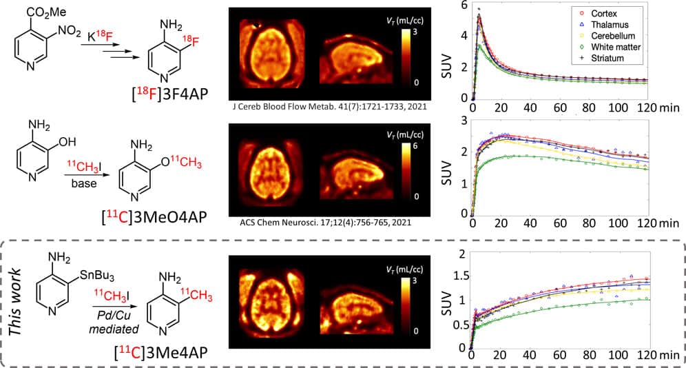
Demyelination, the loss of the insulating sheath of neurons, causes failed or slowed neuronal conduction and contributes to the neurological symptoms in multiple sclerosis, traumatic brain and spinal cord injuries, stroke, and dementia. In demyelinated neurons, the axonal potassium channels Kv1.1 and Kv1.2, generally under the myelin sheath, become exposed and upregulated. Therefore, imaging these channels using positron emission tomography can provide valuable information for disease diagnosis and monitoring. Here, we describe a novel tracer for Kv1 channels, [11C]3-methyl-4-aminopyridine ([11C]3Me4AP). [11C]3Me4AP was efficiently synthesized via Pd(0)–Cu(I) comediated Stille cross-coupling of a stannyl precursor containing a free amino group. Evaluation of its imaging properties in rats and nonhuman primates showed that [11C]3Me4AP has a moderate brain permeability and slow kinetics. Additional evaluation in monkeys showed that the tracer is metabolically stable and that a one-tissue compartment model can accurately model the regional brain time–activity curves. Compared to the related tracers [18F]3-fluoro-4-aminopyridine ([18F]3F4AP) and [11C]3-methoxy-4-aminopyridine ([11C]3MeO4AP), [11C]3Me4AP shows lower initial brain uptake, which indicates reduced permeability to the blood–brain barrier and slower kinetics, suggesting higher binding affinity consistent with in vitro studies. While the slow kinetics and strong binding affinity resulted in a tracer with less favorable properties for imaging the brain than its predecessors, these properties may make 3Me4AP useful as a therapeutic.
Sun Y, et. al., ACS Chem. Neurosci. 2022, 13, 23, 3342–3351
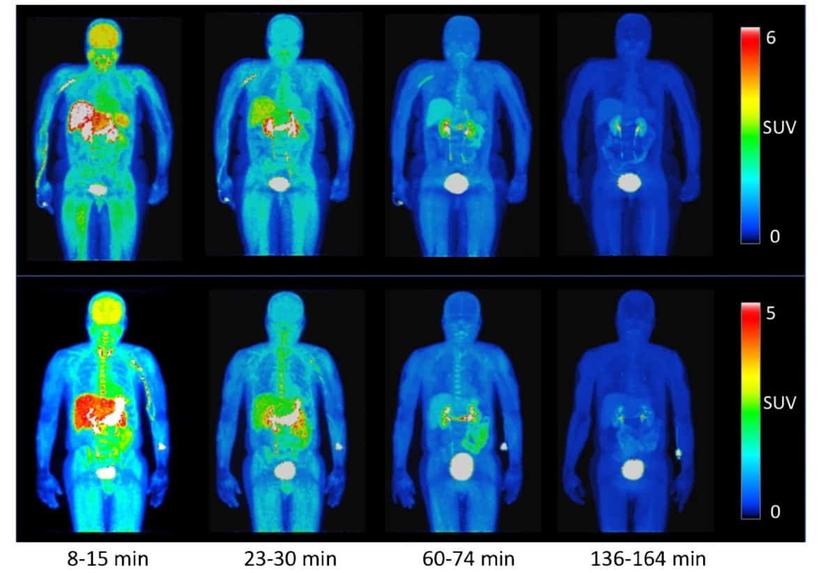
Human biodistribution and radiation dosimetry of the demyelination tracer [18F]3F4AP
Researchers evaluated the safety and effectiveness of a new PET radiotracer called [18F]3F4AP in healthy human volunteers. The radiotracer targets voltage-gated potassium (K+) channels and has shown promise for imaging demyelinated lesions in animal models of neurological diseases, such as multiple sclerosis and dementia. The study found that the radiotracer distributed throughout the body with the highest levels of activity in the kidneys, urinary bladder, stomach, liver, spleen, and brain, and was well-tolerated with no adverse events occurring.
Abstract
Purpose
[18F]3F4AP is a novel PET radiotracer that targets voltage-gated potassium (K+) channels and has shown promise for imaging demyelinated lesions in animal models of neurological diseases. This study aimed to evaluate the biodistribution, safety, and radiation dosimetry of [18F]3F4AP in healthy human volunteers.
Methods
Four healthy volunteers (2 females) underwent a 4-h dynamic PET scan from the cranial vertex to mid-thigh using multiple bed positions after administration of 368 ± 17.9 MBq (9.94 ± 0.48 mCi) of [18F]3F4AP. Volumes of interest for relevant organs were manually drawn guided by the CT, and PET images and time-activity curves (TACs) were extracted. Radiation dosimetry was estimated from the integrated TACs using OLINDA software. Safety assessments included measuring vital signs immediately before and after the scan, monitoring for adverse events, and obtaining a comprehensive metabolic panel and electrocardiogram within 30 days before and after the scan.
Results
[18F]3F4AP distributed throughout the body with the highest levels of activity in the kidneys, urinary bladder, stomach, liver, spleen, and brain and with low accumulation in muscle and fat. The tracer cleared quickly from circulation and from most organs. The clearance of the tracer was noticeably faster than previously reported in nonhuman primates (NHPs). The average effective dose (ED) across all subjects was 12.1 ± 2.2 μSv/MBq, which is lower than the estimated ED from the NHP studies (21.6 ± 0.6 μSv/MBq) as well as the ED of other fluorine-18 radiotracers such as [18F]FDG (~ 20 μSv/MBq). No differences in ED between males and females were observed. No substantial changes in safety assessments or adverse events were recorded.
Conclusion
The biodistribution and radiation dosimetry of [18F]3F4AP in humans are reported for the first time. The average total ED across four subjects was lower than most 18F-labeled PET tracers. The tracer and study procedures were well tolerated, and no adverse events occurred.
Brugarolas P*, Wilks MQ*, et al. Eur J Nuc Med Molec Imag. 2023; 50(2):344-351.
PET imaging studies to investigate functional expression of mGluR2 using [11C]mG2P001
This study aimed to investigate the function of mGluR2, a critical component in treating some neurological and psychiatric conditions, by utilizing PET imaging with [11C]mG2P001, a potent mGluR2 positive allosteric modulator. Results showed that the cooperative binding mechanism between mG2P001 and glutamate can enhance mGluR2 expression in rat brains, as demonstrated by an increase in [11C]mG2P001 accumulation. The imaging technique also proved effective in non-human primates, providing a sensitive biomarker for mGluR2 expression affected by tissue glutamate concentration. These findings may pave the way for further drug development and treatment for brain disorders.

Metabotropic glutamate receptor 2 (mGluR2) has been extensively studied for the treatment of various neurological and psychiatric disorders. Understanding of the mGluR2 function is pivotal in supporting the drug discovery targeting mGluR2. Herein, the positive allosteric modulation of mGluR2 was investigated via the in vivo positron emission tomography (PET) imaging using 2-((4-(2-[11C]methoxy-4-(trifluoromethyl)phenyl)piperidin-1-yl)methyl)-1-methyl-1H-imidazo[4,5-b]pyridine ([11C]mG2P001). Distinct from the orthosteric compounds, pretreatment with the unlabeled mG2P001, a potent mGluR2 positive allosteric modulator (PAM), resulted in a significant increase instead of decrease of the [11C]mG2P001 accumulation in rat brain detected by PET imaging. Subsequent in vitro studies with [3H]mG2P001 revealed the cooperative binding mechanism of mG2P001 with glutamate and its pharmacological effect that contributed to the enhanced binding of [3H]mG2P001 in transfected CHO cells expressing mGluR2.
![Structural MRI (MEMPRAGE) and [11C]mG2P001 Logan VT images for the baseline (middle) and blocking conditions (lower)](https://gordon.mgh.harvard.edu/cmitt/wp-content/uploads/2023/04/images_large_10.1177_0271678x221130387-fig5.jpeg)
The in vivo PET imaging and quantitative analysis of [11C]mG2P001 in non-human primates (NHPs) further validated the characteristics of [11C]mG2P001 as an imaging ligand for mGluR2. Self-blocking studies in primates enhanced accumulation of [11C]mG2P001. Altogether, these studies show that [11C]mG2P001 is a sensitive biomarker for mGluR2 expression and the binding is affected by the tissue glutamate concentration.
Yuan G*, Dhaynaut M*, et. al. J. Med. Chem. 2022, 65, 14, 9939–9954
Design, Synthesis, and Characterization of [18F]mG2P026 as a High-Contrast PET Imaging Ligand for Metabotropic Glutamate Receptor 2
![Structural MRI (MEMPRAGE) and [18F]9 Logan VT images at t*30 min for the baseline and pretreatment condition](https://gordon.mgh.harvard.edu/cmitt/wp-content/uploads/2022/12/images_large_jm2c00593_0007.jpg)
An array of triazolopyridines based on JNJ-46356479 (6) were synthesized as potential positron emission tomography radiotracers for metabotropic glutamate receptor 2 (mGluR2). The selected candidates 8-10 featured enhanced positive allosteric modulator (PAM) activity (20-fold max.) and mGluR2 agonist activity (25-fold max.) compared to compound 6 in the cAMP GloSensor assays. Radiolabeling of compounds 8 and 9 (mG2P026) was achieved via Cu-mediated radiofluorination with satisfactory radiochemical yield, >5% (non-decay-corrected); high molar activity, >180 GBq/μmol; and excellent radiochemical purity, >98%. Preliminary characterization of [18F]8 and [18F]9 in rats confirmed their excellent brain permeability and binding kinetics. Further evaluation of [18F]9 in a non-human primate confirmed its superior brain heterogeneity in mapping mGluR2 and higher affinity than [18F]6. Pretreatment with different classes of PAMs in rats and a primate led to similarly enhanced brain uptake of [18F]9. As a selective ligand, [18F]9 has the potential to be developed for translational studies.
Yuan G, et. al., J Med Chem 2022 65 (14):9939-9954
Radiochemical Synthesis and Evaluation in Non-Human Primates of 3-[11C]methoxy-4-aminopyridine: A Novel PET Tracer for Imaging Potassium Channels in the CNS
![CT and [11C]3MeO4AP parametric images of NHP. Time activity curves showing distinct kinetics in area of injury compared to other regions.](https://gordon.mgh.harvard.edu/cmitt/wp-content/uploads/2022/12/nihms-1682623-f0005.jpg)
Demyelination, the loss of the protecting sheath of neurons, contributes to disability in many neurological diseases. In order to fully understand its role in different diseases and to monitor treatments aiming at reversing this process, it would be valuable to have PET radiotracers that can detect and quantify molecular changes involved in demyelination such as the uncovering and upregulation of the axonal potassium channels Kv1.1 and Kv1.2. Carbon-11 labeled radiotracers present the advantage of allowing for multiple scans on the same subject in the same day. Here, we describe [11C]3MeO4AP, a novel 11C-labeled version of the K+ channel tracer [18F]3F4AP, and characterize its imaging properties in two non-human primates including a monkey with a focal brain injury sustained during a surgical procedure 3 years prior to imaging. Our findings show that [11C]3MeO4AP is brain permeable, metabolically stable and has high plasma availability. When compared with [18F]3F4AP, [11C]3MeO4AP shows very high correlation in volumes of distribution (VT), confirming a common target. [11C]3MeO4AP shows slower washout than [18F]3F4AP, suggesting stronger binding. Finally, similar to [18F]3F4AP, [11C]3MeO4AP is highly sensitive to the focal brain injury. All these features make it a promising radioligand for imaging demyelinated lesions.
Guehl NJ, et. al., ACS Chem Neurosci 2021 12 (4):756-765
Positron annihilation localization by nanoscale magnetization
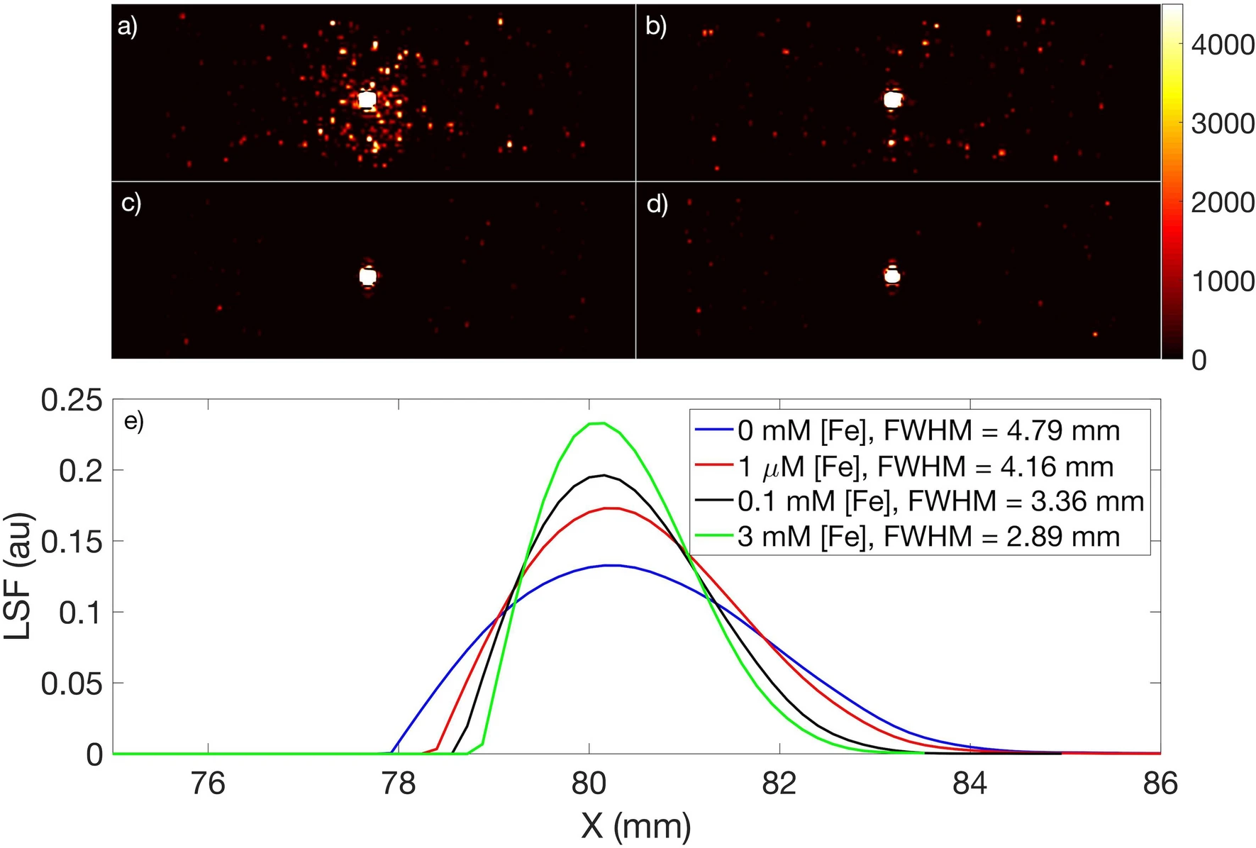
In positron emission tomography (PET), the finite range over which positrons travel before annihilating with an electron places a fundamental physical limit on the spatial resolution of PET images. After annihilation, the photon pair detected by the PET instrumentation is emitted from a location that is different from the positron-emitting source, resulting in image blurring. Here, we report on the localization of positron range, and hence annihilation quanta, by strong nanoscale magnetization of superparamagnetic iron oxide nanoparticles (SPIONs) in PET-MRI. We found that positron annihilations localize within a region of interest by up to 60% more when SPIONs are present (with [Fe] = 3 mM) compared to when they are not. The resulting full width at half maximum of the PET scans showed the spatial resolution improved by up to ≈≈ 30%. We also found evidence suggesting that the radiolabeled SPIONs produced up to a six-fold increase in ortho-positronium. These results may also have implications for emerging cancer theranostic strategies, where charged particles are used as therapeutic as well as diagnostic agents and improved dose localization within a tumor is a determinant of better treatment outcomes.
Y.H. Gholami, et. al., Scientific Reports volume 10, Article number: 20262 (2020)
A Radio-Nano-Platform for T1/T2 Dual-Mode PET-MR Imaging
Purpose: This study aimed to develop a chelate-free radiolabeled nanoparticle platform for simultaneous positron emission tomography (PET) and magnetic resonance (MR) imaging that provides contrast-enhanced diagnostic imaging and significant image quality gain by integrating the high spatial resolution of MR with the high sensitivity of PET.
Methods: A commercially available super-paramagnetic iron oxide nanoparticle (SPION) (Feraheme®, FH) was labeled with the [89Zr]Zr using a novel chelate-free radiolabeling technique, heat-induced radiolabeling (HIR). Radiochemical yield (RCY) and purity (RCP) were measured using size exclusion chromatography (SEC) and radio-thin layer chromatography (radio-TLC). Characterization of the non-radioactive isotope 90Zr-labeled FH was performed by transmission electron microscopy (TEM). Simultaneous PET-MR phantom imaging was performed with different 89Zr-FH concentrations. The MR quantitative image analysis determined the contrast-enhancing properties of FH. The signal-to-noise ratio (SNR) and full-width half-maximum (FWHM) of the line spread function (LSF) were calculated before and after co-registering the PET and MR image data.
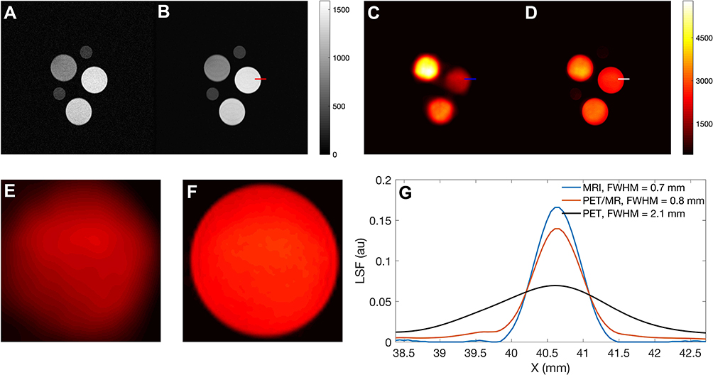
Results: High RCY (92%) and RCP (98%) of the [89Zr]Zr-FH product was achieved. TEM analysis confirmed the 90Zr atoms adsorption onto the SPION surface (≈ 10% average radial increase). Simultaneous PET-MR scans confirmed the capability of the [89Zr]Zr-FH nano-platform for this multi-modal imaging technique. Relative contrast image analysis showed that [89Zr]Zr-FH can act as a dual-mode T1/T2 contrast agent. For co-registered PET-MR images, higher spatial resolution (FWHM enhancement ≈ 3) and SNR (enhancement ≈ 8) was achieved at a clinical dose of radio-isotope and Fe.
Conclusion: Our results demonstrate FH is a highly suitable SPION-based platform for chelate-free labeling of PET tracers for hybrid PET-MR. The high RCY and RCP confirmed the robustness of the chelate-free HIR technique. An overall image quality gain was achieved compared to PET- or MR-alone imaging with a relatively low dosage of [89Zr]Zr-FH. Additionally, FH is suitable as a dual-mode T1/T2 MR image contrast agent.
Y.H. Gholami, et. al., Int J Nanomedicine. 2020 Feb 24;15:1253-1266. doi: 10.2147/IJN.S241971
A Chelate-Free Nano-Platform for Incorporation of Diagnostic and Therapeutic Isotopes
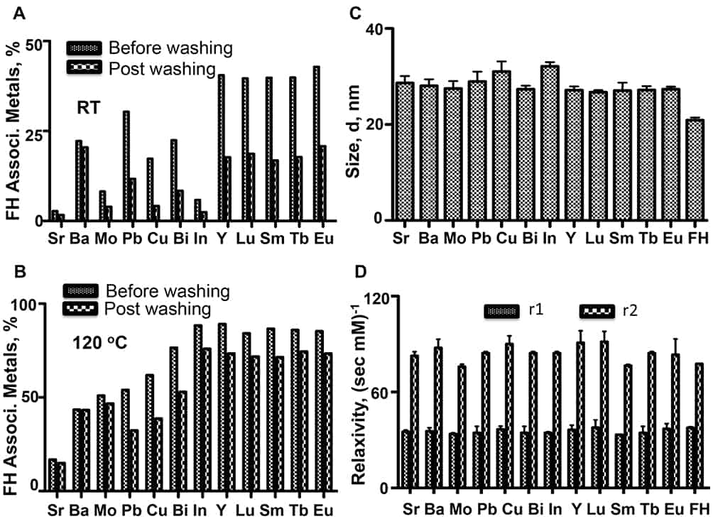
Purpose: Using our chelate-free, heat-induced radiolabeling (HIR) method, we show that a wide range of metals, including those with radioactive isotopologues used for diagnostic imaging and radionuclide therapy, bind to the Feraheme (FH) nanoparticle (NP), a drug approved for the treatment of iron anemia.
Material and methods: FH NPs were heated (120°C) with nonradioactive metals, the resulting metal-FH NPs were characterized by inductively coupled plasma mass spectrometry (ICP-MS), dynamic light scattering (DLS), and r1 and r2 relaxivities obtained by nuclear magnetic relaxation spectrometry (NMRS). In addition, the HIR method was performed with [90Y]Y3+, [177Lu]Lu3+, and [64Cu]Cu2+, the latter with an HIR technique optimized for this isotope. Optimization included modifying reaction time, temperature, and vortex technique. Radiochemical yield (RCY) and purity (RCP) were measured using size exclusion chromatography (SEC) and thin-layer chromatography (TLC).
Results: With ICP-MS, metals incorporated into FH at high efficiency were bismuth, indium, yttrium, lutetium, samarium, terbium and europium (>75% @ 120 oC). Incorporation occurred with a small (less than 20%) but statistically significant increases in size and the r2 relaxivity. An improved HIR technique (faster heating rate and improved vortexing) was developed specifically for copper and used with the HIR technique and [64Cu]Cu2+. Using SEC and TLC analyses with [90Y]Y3+, [177Lu]Lu3+ and [64Cu]Cu2+, RCYs were greater than 85% and RCPs were greater than 95% in all cases.
Conclusion: The chelate-free HIR technique for binding metals to FH NPs has been extended to a range of metals with radioisotopes used in therapeutic and diagnostic applications. Cations with f-orbital electrons, more empty d-orbitals, larger radii, and higher positive charges achieved higher values of RCY and RCP in the HIR reaction. The ability to use a simple heating step to bind a wide range of metals to the FH NP, a widely available approved drug, may allow this NP to become a platform for obtaining radiolabeled nanoparticles in many settings.
Y.H. Gholami, et. al. Int J Nanomedicine. 2020 Jan 7;15:31-47. doi: 10.2147/IJN.S227931.
Dependence of fluorodeoxyglucose (FDG) uptake on cell cycle and dry mass: a single-cell study using a multi-modal radiography platform
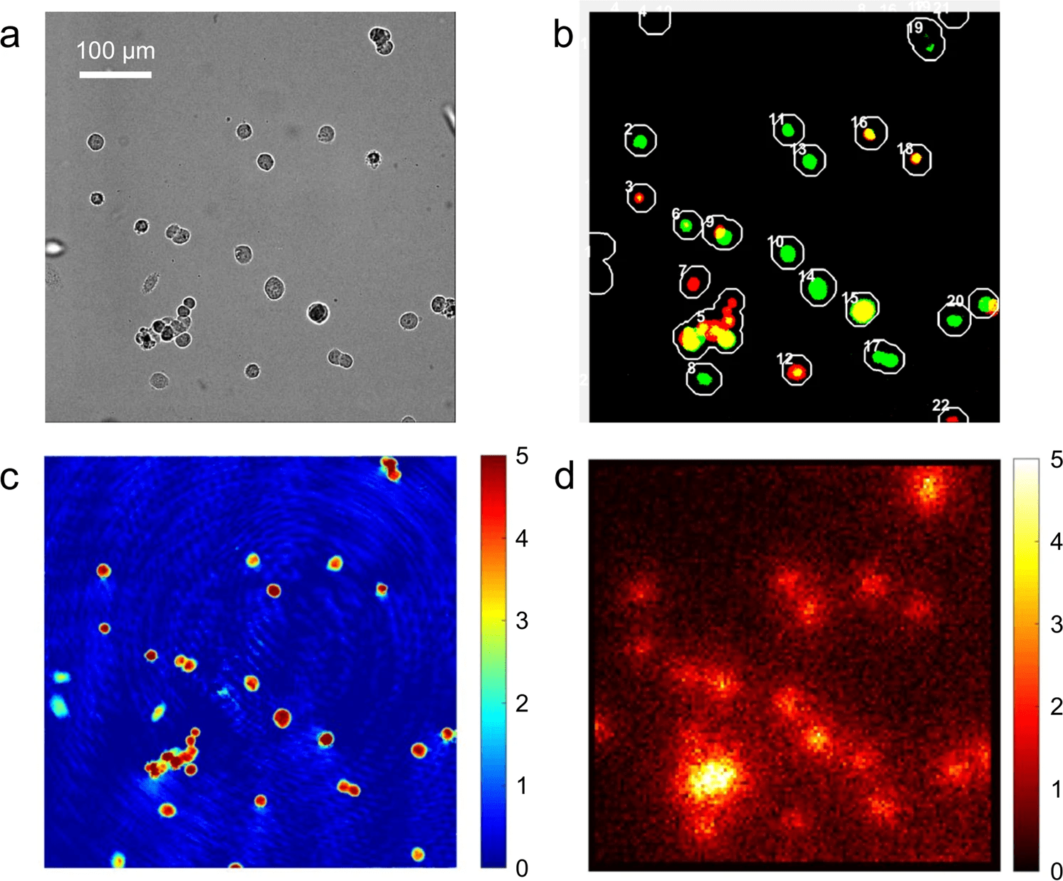
High glucose uptake by cancer compared to normal tissues has long been utilized in fluorodeoxyglucose-based positron emission tomography (FDG-PET) as a contrast mechanism. The FDG uptake rate has been further related to the proliferative potential of cancer, specifically the proliferation index (PI) – the proportion of cells in S, G2 or M phases.
The underlying hypothesis was that the cells preparing for cell division would consume more energy and metabolites as building blocks for biosynthesis. Despite the wide clinical use, mixed reports exist in the literature on the relationship between FDG uptake and PI. This may be due to the large variation in cancer types or methods adopted for the measurements. Of note, the existing methods can only measure the average properties of a tumor mass or cell population with highly-heterogeneous constituents.
In this study, we have built a multi-modal live-cell radiography system and measured the [18F]FDG uptake by single HeLa cells together with their dry mass and cell cycle phase. The results show that HeLa cells take up twice more [18F]FDG in S, G2 or M phases than in G1 phase, which confirms the association between FDG uptake and PI at a single-cell level. Importantly, we show that [18F]FDG uptake and cell dry mass have a positive correlation in HeLa cells, which suggests that high [18F]FDG uptake in S, G2 or M phases can be largely attributed to increased dry mass, rather than the activities preparing for cell division. This interpretation is consistent with recent observations that the energy required for the preparation of cell division is much smaller than that for maintaining house-keeping proteins.
Y. Sung, et. al., Scientific Reports volume 10, Article number: 4280 (2020)
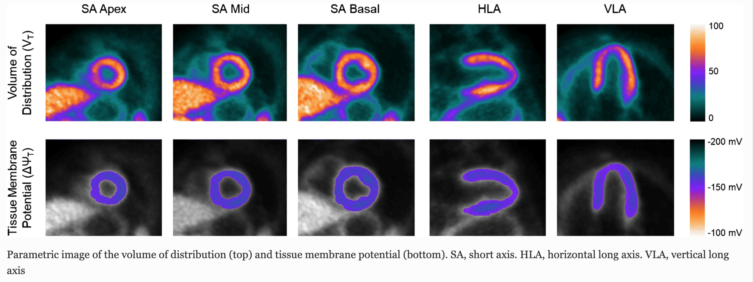
Quantitative in vivo mapping of myocardial mitochondrial membrane potential
Alteration in mitochondrial membrane potential (ΔΨm) is an important feature of many pathologic processes, including heart failure, cardiotoxicity, ventricular arrhythmia, and myocardial hypertrophy.
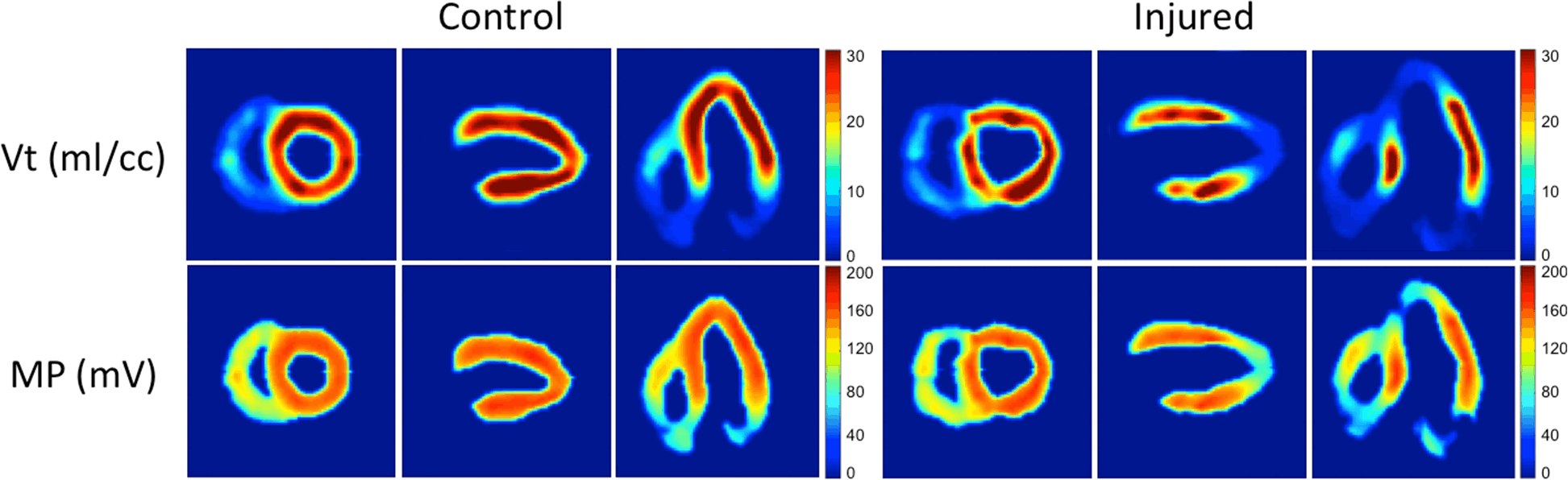
We used a swine cardiac model to validate a method for simplified measurement of total membrane potential. A bolus-plus-infusion (B/I) of [18F]-triphenylphosphonium ([18F]TPP+) was imaged dynamically and used to measure and map the total membrane potential, ΔΨT, as the sum of ΔΨm and cellular (ΔΨc) electrical potentials.
We derived equations expressing ΔΨT as a function of VT and the volume-fractions of mitochondria and fECS. Seventeen segment polar maps and parametric images of ΔΨT were calculated in millivolts (mV).
In controls, mean segmental ΔΨT = -129.4±1.4 mV (SEM). In pigs with segmental tissue injury, ΔΨT was clearly separated from control segments but variable, in the range -100 to 0 mV. The quality of ΔΨT maps was excellent, with low noise and good resolution. Measurements of ΔΨT in the left ventricle of pigs agree with previous in in-vitro measurements. These studies demonstrate the first in vivo application of quantitative mapping of total tissue membrane potential, ΔΨT.
NM Alpert, et. al., PLoS One. 2018 Jan 16;13(1):e0190968.
In-vivo Imaging of Mitochondrial Depolarization of Myocardium With Positron Emission Tomography and a Proton Gradient Uncoupler
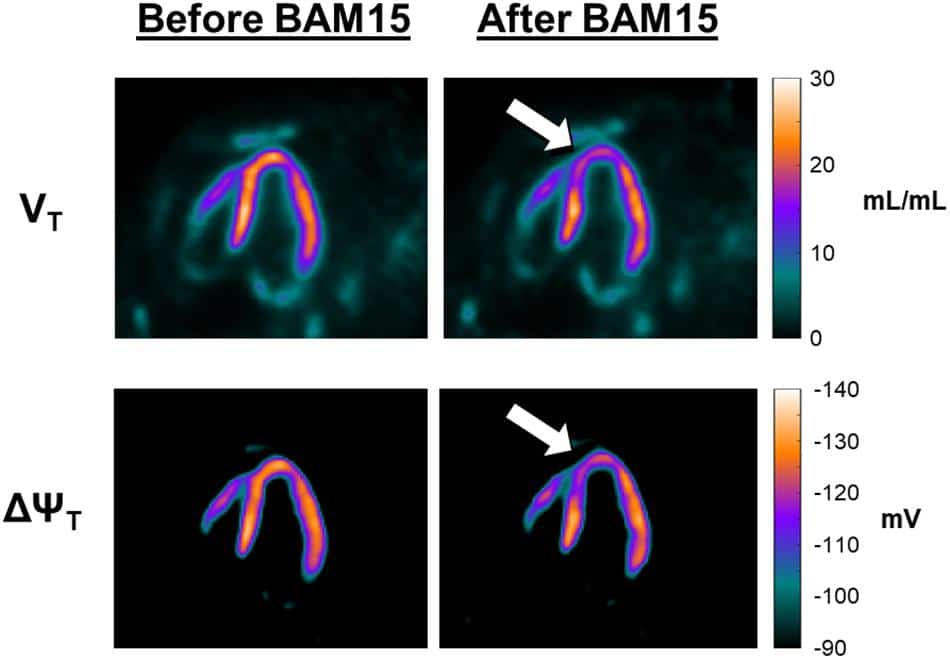
Background: We recently reported a method using positron emission tomography (PET) and the tracer 18F-labeled tetraphenylphosphonium (18F-TPP+) for mapping the tissue (i.e., cellular plus mitochondrial) membrane potential (ΔΨT) in the myocardium. The purpose of this work is to provide additional experimental evidence that our methods can be used to observe transient changes in the volume of distribution for 18F-TPP+ and mitochondrial membrane potential (ΔΨm).
Methods: We tested these hypotheses by measuring decreases of 18F-TPP+ concentration elicited when a proton gradient uncoupler, BAM15, is administered by intracoronary infusion during PET scanning. BAM15 is the first proton gradient uncoupler shown to affect the mitochondrial membrane without affecting the cellular membrane potential. Preliminary dose response experiments were conducted in two pigs to determine the concentration of BAM15 infusate necessary to perturb the 18F-TPP+ concentration. More definitive experiments were performed in two additional pigs, in which we administered an intravenous bolus plus infusion of 18F-TPP+ to reach secular equilibrium followed by an intracoronary infusion of BAM15.
Results: Intracoronary BAM15 infusion led to a clear decrease in 18F-TPP+ concentration, falling to a lower level, and then recovering. A second BAM15 infusion reduced the 18F-TPP+ level in a similar fashion. We observed a maximum depolarization of 10 mV as a result of the BAM15 infusion.
Summary: This work provides evidence that the total membrane potential measured with 18F-TPP+ PET is sensitive to temporal changes in mitochondrial membrane potential.
NM Alpert, et. al. Front Physiol. 2020 May 15;11:491. doi: 10.3389/fphys.2020.00491.
In vivo quantitative mapping of human mitochondrial cardiac membrane potential: a feasibility study
We have made an in vivo, non-invasive, assessment of regional ΔΨm in the myocardium of normal human subjects using dynamic imaging on a PET/MR scanner during and after a bolus+infusion of [18F]TPP+. The extracellular space fraction (fECS) was simultaneously measured using MR T1-mapping images acquired at baseline and 15 min after gadolinium injection.
The tissue membrane potential (ΔΨT), a proxy of ΔΨm, was calculated from the myocardial tracer concentration at secular equilibrium, blood concentration, and fECS measurements using a model based on the Nernst equation.
Average ΔΨT was in excellent agreement with previous in vitro assessment. This feasibility study lays the foundation for further investigations to assess these potential role of PET/MR in in vivo quantification of the mitochondrial function, and has the potential to provide new diagnostic and prognostic information for several cardiac diseases as well as allowing therapy monitoring.
M Pelletier-Galarneau, et. al. Eur J Nucl Med Mol Imaging. 2020 Jul 27.
Synthesis and Characterization of Fluorine-18-Labeled N-(4-Chloro-3-((fluoromethyl- d2)thio)phenyl)picolinamide for Imaging of mGluR4 in Brain
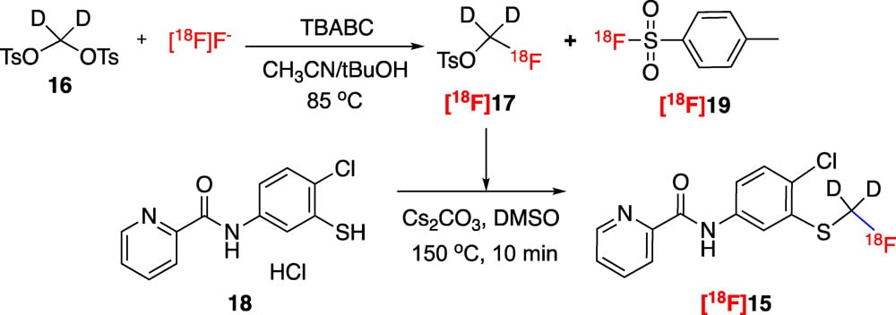
We have synthesized and characterized [18F]-N-(4-chloro-3-((fluoromethyl-d2)thio)phenyl)-picolinamide ([18F]15) as a potential ligand for the positron emission tomography (PET) imaging of mGluR4 in the brain. Radioligand [18F]15 displays central nervous system drug-like properties, including mGluR4 affinity, potent mGluR4 PAM activity, and selectivity against other mGluRs, as well as sufficient metabolic stability. Radiosynthesis was carried out in two steps. The radiochemical yield of [18F]15 was 11.6 ± 2.9% (n = 7, decay corrected) with a purity of 99% and a molar activity of 84.1 ± 11.8 GBq/μmol. Ex vivo biodistribution studies showed reversible binding of [18F]15 in all investigated tissues including the brain, liver, heart, lungs, and kidneys.
PET imaging studies in male Sprague Dawley rats showed that [18F]15 accumulates in the brain regions known to express mGluR4. Pretreatment with the unlabeled mGluR4 PAM compounds 13 (methylthio analogue) and 15 showed significant dose-dependent blocking effects. These results suggest that [18F]15 is a promising radioligand for PET imaging mGluR4 in the brain.
J. Wang, J Med Chem. 2020 Mar 26;63(6):3381-3389.
Evaluation of pharmacokinetic modeling strategies for in-vivo quantification of tau with the radiotracer [F]MK6240 in human subjects
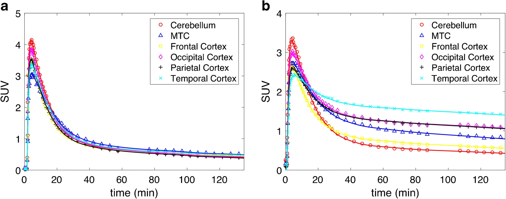
Purpose: [18F]MK6240 was developed for PET imaging of tau aggregates, which are implicated in Alzheimer’s disease. The goal of this work was to evaluate the kinetics of [18F]MK6240 and to investigate different strategies for in-vivo quantification of tau aggregates in humans.
Methods: Thirty-five subjects, consisting of 18 healthy controls (CTRL), 11 subjects with mild cognitive impairment (MCI) and six with Alzheimer’s Disease (AD), underwent dynamic [18F]MK6240 PET scans. Arterial blood measurements were collected in 16 subjects (eight CTRLs, six MCIs and two AD) to measure whole blood and plasma concentration time courses. Radiometabolite analysis was performed on a subset of plasma samples. Various compartmental model configurations as well as the Logan and multilinear analysis (MA1) graphical methods with arterial plasma input function were tested. Simplified reference tissue methods were investigated, including Logan distribution volume ratio (DVR), multilinear reference tissue method (MRTM2), and static SUV ratio using the cerebellum as a reference region.
Results: Whole blood:plasma ratio stabilized to 0.66 ± 0.01 after 15 min. Percent parent in plasma (%PP) followed a single exponential and ranged from 0 to 10% at 90 min. [18F]MK6240 in gray matter peaked quickly (SUV > 2 at ~3 min). The preferred compartmental model was a reversible two-tissue compartment model, with the blood contribution included as a model parameter (2T4k1v). Compartmental and graphical analysis methods with arterial input functions yielded concordant results, but rapid metabolism raised challenges for blood-based quantification. MCI and AD subjects demonstrated a broad range of VT as compared to CTRL subjects. DVR from MRTM2 and Logan reference tissue methods correlated with DVR calculated indirectly from compartmental modeling, but underestimation was observed in data sets with very high binding (DVR > 3). SUVR also underestimated indirect DVR from blood-based analyses in high binding regions, although a non-linear relationship was exhibited.
Conclusions: [18F]MK6240 exhibited a wide dynamic range of uptake, with binding patterns in MCI/AD subjects consistent with neurofibrillary tau deposition patterns. Linearized reference tissue methods using an estimated average tissue-to-plasma efflux constant [Formula: see text] and static SUVR agreed well with blood-based methods for most data sets; however, discrepancies were noted in the highest binding cases. Caution should therefore be exercised in application of simplified methods to such data sets, and in quantitative interpretation of corresponding outcomes.
NJ Guehl, et. al. Eur J Nucl Med Mol Imaging. 2019 Sep;46(10):2099-2111. doi: 10.1007/s00259-019-04419-z.
Assessment of Striatal Dopamine Transporter Binding in Individuals With Major Depressive Disorder: In Vivo Positron Emission Tomography and Postmortem Evidence
Importance: Major depressive disorder (MDD) might involve dopamine (DA) reductions. The DA transporter (DAT) regulates DA clearance and neurotransmission and is sensitive to DA levels, with preclinical studies (including those involving inescapable stressors) showing that DAT density decreases when DA signaling is reduced. Despite preclinical data, evidence of reduced DAT in MDD is inconclusive.
Objective: Using a highly selective DAT positron emission tomography (PET) tracer ([11C] altropane), DAT availability was probed in individuals with MDD who were not taking medication. Levels of DAT expression were also evaluated in postmortem tissues from donors with MDD who died by suicide.
Design, setting, and participants: This cross-sectional PET study was conducted at McLean Hospital (Belmont, Massachusetts) and Massachusetts General Hospital (Boston) and enrolled consecutive individuals with MDD who were not taking medication and demographically matched healthy controls between January 2012 and March 2014. Brain tissues were obtained from the Douglas-Bell Canada Brain Bank. For the PET component, 25 individuals with current MDD who were not taking medication and 23 healthy controls recruited from McLean Hospital were included (all provided usable data). For the postmortem component, 15 individuals with depression and 14 healthy controls were considered.
Intervention: PET scan.
Main outcomes and measures: Striatal and midbrain DAT binding potential was assessed. For the postmortem component, tyrosine hydroxylase and DAT levels were evaluated using Western blots.

Results: Compared with 23 healthy controls (13 women [56.5%]; mean [SD] age, 26.49 [7.26] years), 25 individuals with MDD (19 women [76.0%]; mean [SD] age, 26.52 [5.92] years) showed significantly lower in vivo DAT availability in the bilateral putamen and ventral tegmental area (Cohen d range, -0.62 to -0.71), and both reductions were exacerbated with increasing numbers of depressive episodes. Unlike healthy controls, the MDD group failed to show an age-associated reduction in striatal DAT availability, with young individuals with MDD being indistinguishable from older healthy controls. Moreover, DAT availability in the ventral tegmental area was lowest in individuals with MDD who reported feeling trapped in stressful circumstances. Lower DAT levels (and tyrosine hydroxylase) in the putamen of MDD compared with healthy controls were replicated in postmortem analyses (Cohen d range, -0.92 to -1.15).
Conclusions and relevance: Major depressive disorder, particularly with recurring episodes, is characterized by decreased striatal DAT expression, which might reflect a compensatory downregulation due to low DA signaling within mesolimbic pathways.
DA Pizzagalli, et. al. JAMA Psychiatry. 2019 Aug 1;76(8):854-861. doi: 10.1001/jamapsychiatry.2019.0801.
Autoradiography validation of novel tau PET tracer [F-18]-MK-6240 on human postmortem brain tissue
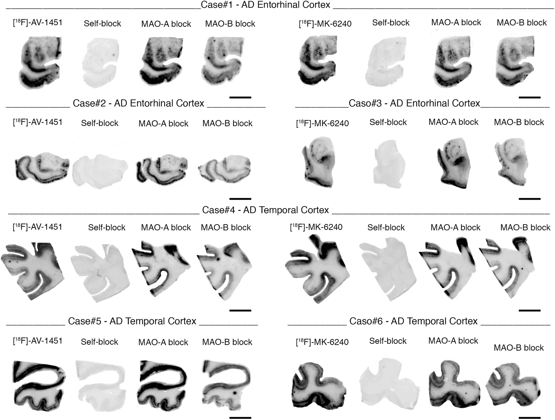
[F-18]-MK-6240, a novel tau positron emission tomography (PET) tracer recently discovered for the in vivo detection of neurofibrillary tangles, has the potential to improve diagnostic accuracy in the detection of Alzheimer disease. We have examined regional and substrate-specific binding patterns as well as possible off-target binding of this tracer on human brain tissue to advance towards its validation. We applied [F-18]-MK-6240 phosphor screen and high resolution autoradiography to postmortem samples from patients with a definite pathological diagnosis of Alzheimer disease, frontotemporal lobar degeneration-tau (Pick’s disease, progressive supranuclear palsy and corticobasal degeneration), chronic traumatic encephalopathy, frontotemporal lobar degeneration-Tar DNA-binding protein 43 (TDP-43), dementia with Lewy bodies, cerebral amyloid angiopathy and elderly controls free of pathologic changes of neurodegenerative disease. We also directly compared the binding properties of [F-18]-MK-6240 and [F-18]-AV-1451 in human tissue, and examined potential nonspecific binding of both tau tracers to monoamine oxidases (MAO) by using autoradiography in the presence of selective monoamine oxidase A (MAO-A) and monoamine oxidase B (MAO-B) inhibitors.
Our data indicate that MK-6240 strongly binds to neurofibrillary tangles in Alzheimer disease but does not seem to bind to a significant extent to tau aggregates in non-Alzheimer tauopathies, suggesting that it may have a limited utility for the in vivo detection of these pathologies. There is no evidence of binding to lesions containing β-amyloid, α-synuclein or TDP-43. In addition, we identified MK-6240 strong off-target binding to neuromelanin and melanin-containing cells, and some weaker binding to areas of hemorrhage. These binding patterns are nearly identical to those previously reported by our group and others for [F-18]-AV-1451. Of note, [F-18]-MK-6240 and [F-18]-AV-1451 autoradiographic binding signals were only weakly displaced by competing concentrations of selective MAO-B inhibitor deprenyl but not by MAO-A inhibitor clorgyline, suggesting that MAO enzymes do not appear to be a significant binding target of any of these two tracers. Together these novel findings provide relevant insights for the correct interpretation of in vivo [F-18]-MK-6240 PET imaging.
C Aguero, et. al. Acta Neuropathol Commun. 2019 Mar 11;7(1):37. doi: 10.1186/s40478-019-0686-6.
Heat-induced radiolabeling and fluorescence labeling of Feraheme nanoparticles for PET/SPECT imaging and flow cytometry
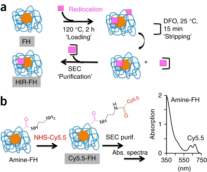
Feraheme (FH) nanoparticles (NPs) have been used extensively for treatment of iron anemia (due to their slow release of ionic iron in acidic environments). In addition, injected FH NPs are internalized by monocytes and function as MRI biomarkers for the pathological accumulation of monocytes in disease.
We have recently expanded these applications by radiolabeling FH NPs for positron emission tomography (PET) or single-photon emission computed tomography (SPECT) imaging using a heat-induced radiolabeling (HIR) strategy. Imaging FH NPs using PET/SPECT has important advantages over MRI due to lower iron doses and improved quantitation of tissue NP concentrations. HIR of FH NPs leaves the physical and biological properties of the NPs unchanged and allows researchers to build on the extensive knowledge obtained about the pharmacokinetic and safety aspects of FH NPs.
In this protocol, we present the step-by-step procedures for heat (120 °C)-induced bonding of three widely employed radiocations (89Zr4+ or 64Cu2+ for PET, and 111In3+ for SPECT) to FH NPs using a chelateless radiocation surface adsorption (RSA) approach. In addition, we describe the conversion of FH carboxyl groups into amines and their reaction with an N-hydroxysuccinimide (NHS) of a Cy5.5 fluorophore. This yields Cy5.5-FH, a fluorescent FH that enables the cells internalizing Cy5.5-FH to be examined using flow cytometry. Finally, we describe procedures for in vivo and ex vivo uptake of Cy5.5-FH by monocytes and for in vivo microPET/CT imaging of HIR-FH NPs. Synthesis of HIR-FH requires experience with working with radioactive cations and can be completed within <4 h. Synthesis of Cy5.5-FH NPs takes ∼17 h.
H Yuan, et. al. Nat Protoc. 2018 Feb;13(2):392-412. doi: 10.1038/nprot.2017.133.
Structural tract alterations predict downstream tau accumulation in amyloid-positive older individuals
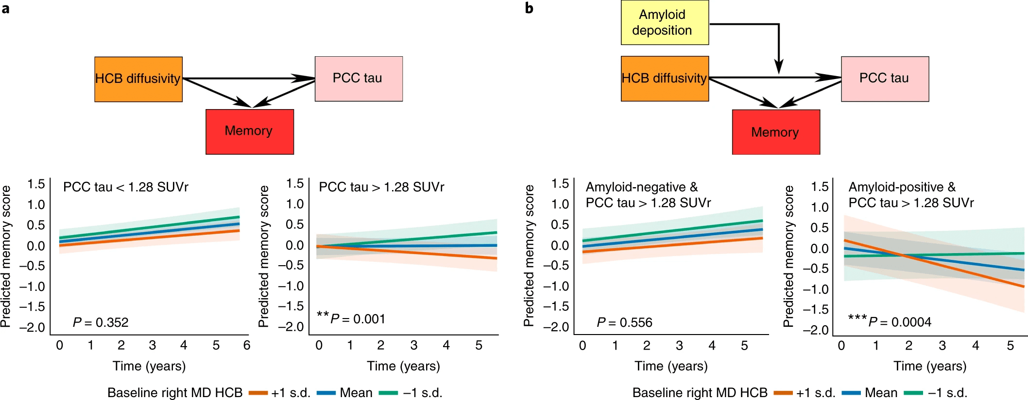
Animal models of Alzheimer’s disease have suggested that tau pathology propagation, facilitated by amyloid pathology, may occur along connected pathways. To investigate these ideas in humans, we combined amyloid scans with longitudinal data on white matter connectivity, hippocampal volume, tau positron emission tomography and memory performance in 256 cognitively healthy older individuals. Lower baseline hippocampal volume was associated with increased mean diffusivity of the connecting hippocampal cingulum bundle (HCB). HCB diffusivity predicted tau accumulation in the downstream-connected posterior cingulate cortex in amyloid-positive but not in amyloid-negative individuals. Furthermore, HCB diffusivity predicted memory decline in amyloid-positive individuals with high posterior cingulate cortex tau binding. Our results provide in vivo evidence that higher amyloid pathology strengthens the association between HCB diffusivity and tau accumulation in the downstream posterior cingulate cortex and facilitates memory decline. This confirms amyloid’s crucial role in potentiating neural vulnerability and memory decline marking the onset of preclinical Alzheimer’s disease.
HIL Jacobs, et. al. Nat Neurosci. 2018 Mar;21(3):424-431. doi: 10.1038/s41593-018-0070-z.
Neurogenetic contributions to amyloid beta and tau spreading in the human cortex
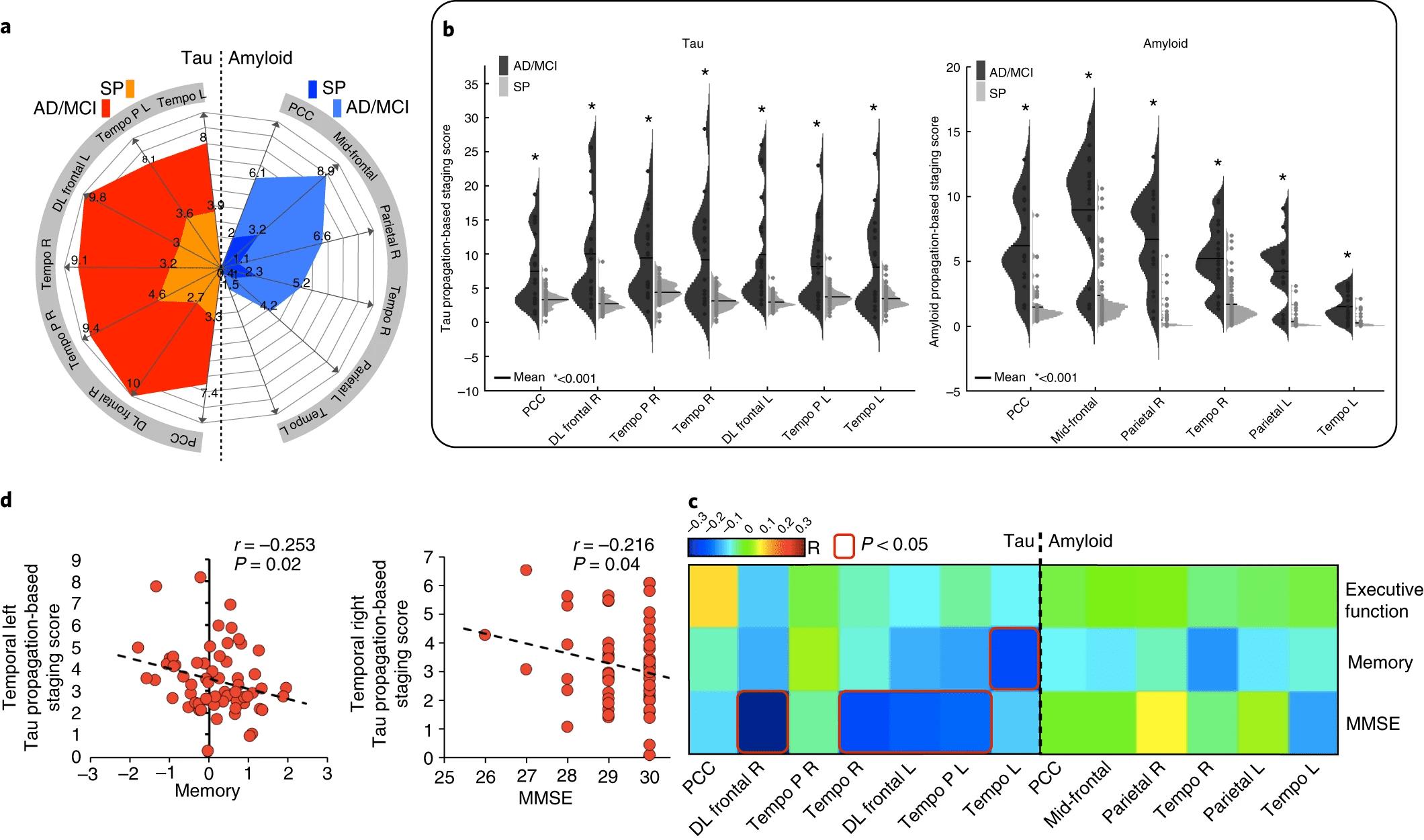
Tau and amyloid beta (Aβ) proteins accumulate along neuronal circuits in Alzheimer’s disease. Unraveling the genetic background for the regional vulnerability of these proteinopathies can help in understanding the mechanisms of pathology progression. To that end, we developed a novel graph theory approach and used it to investigate the intersection of longitudinal Aβ and tau positron emission tomography imaging of healthy adult individuals and the genetic transcriptome of the Allen Human Brain Atlas. We identified distinctive pathways for tau and Aβ accumulation, of which the tau pathways correlated with cognitive levels. We found that tau propagation and Aβ propagation patterns were associated with a common genetic profile related to lipid metabolism, in which APOE played a central role, whereas the tau-specific genetic profile was classified as ‘axon related’ and the Aβ profile as ‘dendrite related’. This study reveals distinct genetic profiles that may confer vulnerability to tau and Aβ in vivo propagation in the human brain.
J Sepulcre, et. al. Nat Med. 2018 Dec;24(12):1910-1918. doi: 10.1038/s41591-018-0206-4