Petibon Y, Sun T, Han PK, Ma C, El Fakhri G, Ouyang J
Phys Med Biol 2019; 64(19):195009
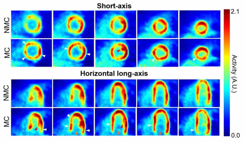
Motion of the myocardium deteriorates the quality and quantitative accuracy of cardiac PET images. We present a method for MR-based cardiac and respiratory motion correction of cardiac PET data and evaluate its impact on estimation of activity and kinetic parameters in human subjects. Three healthy subjects underwent simultaneous dynamic 18F-FDG PET and MRI on a hybrid PET/MR scanner. A cardiorespiratory motion field was determined for each subject using navigator, tagging and golden-angle radial MR acquisitions. Acquired coincidence events were binned into cardiac and respiratory phases using electrocardiogram and list mode-driven signals, respectively. Dynamic PET images were reconstructed with MR-based motion correction (MC) and without motion correction (NMC). Parametric images of 18F-FDG consumption rates (Ki) were estimated using Patlak’s method for both MC and NMC images. MC alleviated motion artifacts in PET images, resulting in improved spatial resolution, improved recovery of activity in the myocardium wall and reduced spillover from the myocardium to the left ventricle cavity. Significantly higher myocardium contrast-to-noise ratio and lower apparent wall thickness were obtained in MC versus NMC images. Likewise, parametric images of Ki calculated with MC data had improved spatial resolution as compared to those obtained with NMC. Consistent with an increase in reconstructed activity concentration in the frames used during kinetic analyses, MC led to the estimation of higher Ki values almost everywhere in the myocardium, with up to 18% increase (mean across subjects) in the septum as compared to NMC. This study shows that MR-based motion correction of cardiac PET results in improved image quality that can benefit both static and dynamic studies.