Dr. Johnson’s Lab has reported on recent work in using Positron emission tomography (PET) to image tau protein deposition in the brain in Alzheimer’s disease (AD).
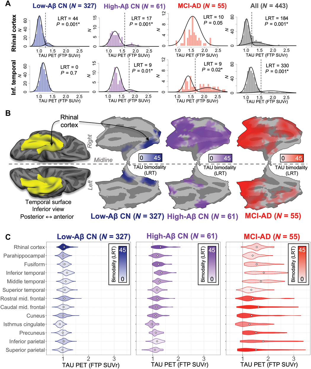
Sanchez et al. applied an automated method to quantify tau progression in patients with AD at different stages. The authors found that tau signal emerges first in the rhinal cortex independently of Aβ. Subsequent tau elevation in the temporal neocortex was associated with age, Aβ, and APOEe4 status. Longitudinal data in a subgroup of patients confirmed the tau trajectory and showed that baseline rhinal cortex tau was highly predictive of subsequent tau spread. Targeting tau in the temporal lobe might slow tau spread and disease progression in patients with AD.
A summary of the article is featured on New Medical
Sanchez, J.S., et al. (2021) The cortical origin and initial spread of medial temporal tauopathy in Alzheimer’s disease assessed with positron emission tomography. Science Translational Medicine. doi.org/10.1126/scitranslmed.abc0655.