Image Reconstruction Technique Provides Unprecedented View of Swallowing and Speech
01/22/2016
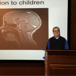
MRI provides several imaging contrasts to study the dynamic muscle movements and functional neural control of speech and swallowing. Leveraging a recently developed low rank imaging approach called partial separability, Dr. Brad Sutton from the University of Illinois at Urbana-Champaign was able to achieve 150 frames per second and provide 3D dynamic imaging of the full vocal tract during speech.The result is an unprecedented view of articulation associated with specific speech sounds.
Brad Sutton is the Associate Professor of Bioengineering and Technical Director of the Biomedical Imaging Center at the University of Illinois at Urbana-Champaign. He was the guest speaker at a clinical research seminar organized by the Gordon Center and hosted by the Massachusetts General Hospital.

Molecular Imaging Pushes Boundaries of Personalized Medicine
01/20/2016
Researchers at the Gordon Center for Medical Imaging are using molecular imaging and nuclear medicine technology to push the boundaries of personalized medicine. We talked to Dr. Rachid Akiki, MD, who is working closely with Dr. Marc Normandin, PhD, in the Gordon Center of the Radiology Department, to find out how.
What is molecular imaging?
Molecular imaging provides detailed pictures of what’s happening inside the body at the smallest level, cells and molecules. Using tracers to illuminate cellular activity, molecular imaging can reveal how a cell behaves and help us understand disease progression. Molecular imaging can confirm a diagnosis, monitor and assess the effectiveness of a treatment like chemotherapy. It is the basis upon which pharmacological companies create new drugs.
How does molecular imaging help you research disease?
We commonly use MRI, CT and PET to take a closer look at the behavior of cells affected by diseases such as cancer, diabetes, heart and kidney problems. Using a small amount of PET tracer, molecular imaging can help improve the ways we diagnose and treat a disease on a case-by-case basis. Cancer cells, for instance, can form unique structures and behave differently from patient to patient.
Tell us about a recent project?
Tell us about yourself?
I am 26 years old and have studied physics at AUB, medicine and medical imaging at BU. Currently I work as a postdoctoral research fellow at Massachusetts General Hospital with an appointment at Harvard medical school on nuclear medicine research topics and their applications in medicine. I like very much my research work and I hope I can affect positively the lives of people significantly while advancing the science. I look forward to start my clinical residency in 2016 and translate my research work to the clinical setting.This article was originally posted by Massachusetts General Hospital Imaging on Wednesday, January 20, 2016