In oncology and cardiology, lesion and defect detection is critical for the diagnosis and treatment of patients. More information about our efforts to improve the medical imaging of lesions in the lungs and the liver, and the detection of heart defects can be found in the links below.
Myocardial Defect Detection
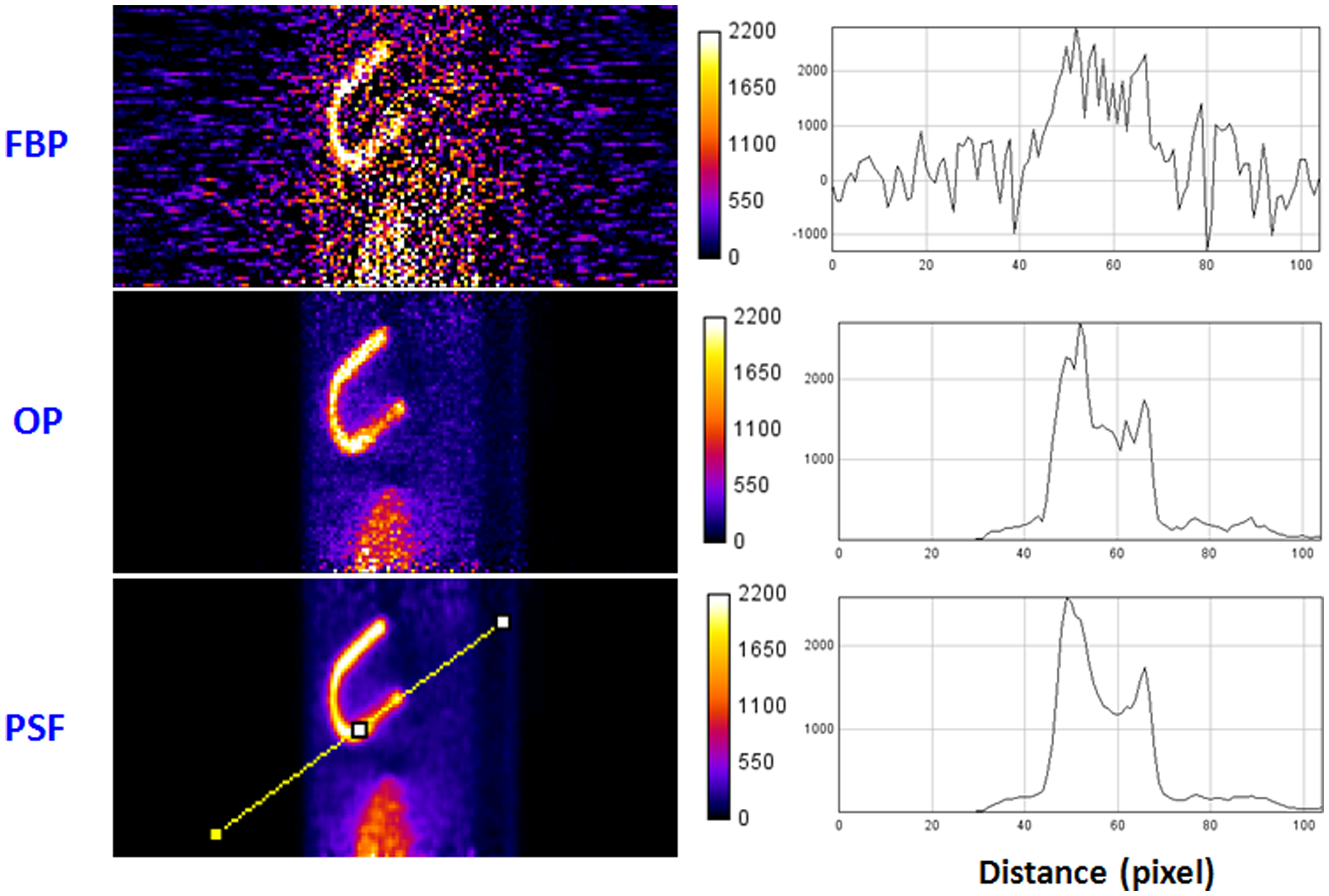
It is expected that both noise and activity distribution can have impact on the detectability of a myocardial defect in a cardiac PET study. We performed phantom studies to investigate the detectability of a defect in the myocardium for different noise levels and activity distributions. We evaluated the performance of three reconstruction schemes: Filtered Back-Projection (FBP), Ordinary Poisson Ordered Subset Expectation Maximization (OP–OSEM), and Point Spread Function corrected OSEM (PSF–OSEM). We used the Channelized Hotelling Observer (CHO) for the task of myocardial defect detection. We found that the detectability of a myocardial defect is almost entirely dependent on the noise level and the contrast between the defect and its surroundings.
Eugene S. Mananga, Georges El Fakhri, Joshua Schaefferkoetter, Ali A. Bonab, Jinsong Ouyang, Myocardial Defect Detection Using PET-CT: Phantom Studies, PLoS ONE, 2014.