Alpert NM, Pelletier-Galarneau M, Kim SJW, Petibon Y, Sun T, Ramos-Torres KM, Normandin MD, El Fakhri G.
Front Physiol. 2020 May 15;11:491.
The purpose of this work is to provide additional experimental evidence that our methods can be used to observe transient changes in the volume of distribution for 18F-TPP+ and mitochondrial membrane potential (ΔΨm).
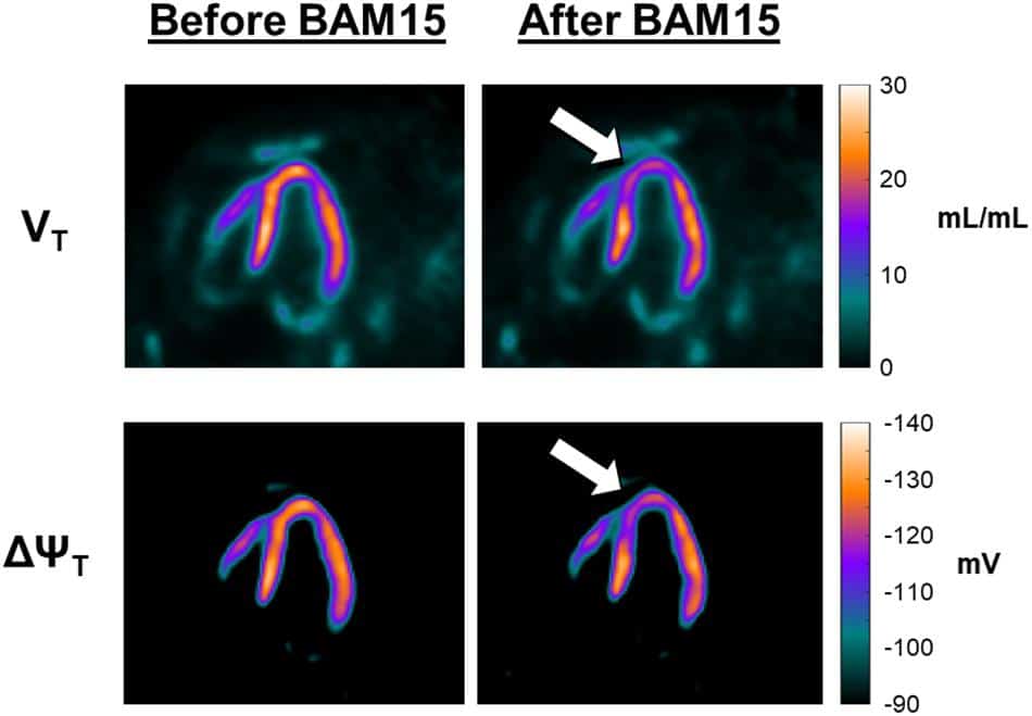
We tested these hypotheses by measuring decreases of 18F-TPP+ concentration elicited when a proton gradient uncoupler, BAM15, is administered by intracoronary infusion during PET scanning. BAM15 is the first proton gradient uncoupler shown to affect the mitochondrial membrane without affecting the cellular membrane potential.
Experiments were conducted in pigs, first to determine the concentration of BAM15 infusate necessary to perturb the 18F-TPP+ concentration, then to administer an intravenous bolus plus infusion of 18F-TPP+ to reach secular equilibrium followed by an intracoronary infusion of BAM15.
Intracoronary BAM15 infusion led to a clear decrease in 18F-TPP+ concentration, falling to a lower level, and then recovering. A second BAM15 infusion reduced the 18F-TPP+ level in a similar fashion. We observed a maximum depolarization of 10 mV as a result of the BAM15 infusion.
This work provides evidence that the total membrane potential measured with 18F-TPP+ PET is sensitive to temporal changes in mitochondrial membrane potential.