Research Interests
The Sepulcre lab focuses on brain imaging studies aiming at the understanding of large-scale brain networks implicated in human cognition and neurodegenerative disorders. We also devote a substantial part of our work developing cutting-edge network methodologies for different brain imaging modalities such as functional connectivity MRI and PET imaging.
Our specific research interest aligns with the following topics:
Our specific research interest aligns with the following topics:
- Systems Neuroscience
- Brain Functional Streams
- Brain Hierarchical Organization
- Multimodal Sensory Integration
- Language Neuroarchitecture
- Neurodegenerative Disorders
Results from articles
Stepwise Connectivity of the Sensory Cortex Reveals the Multimodal Organization of the Human Brain
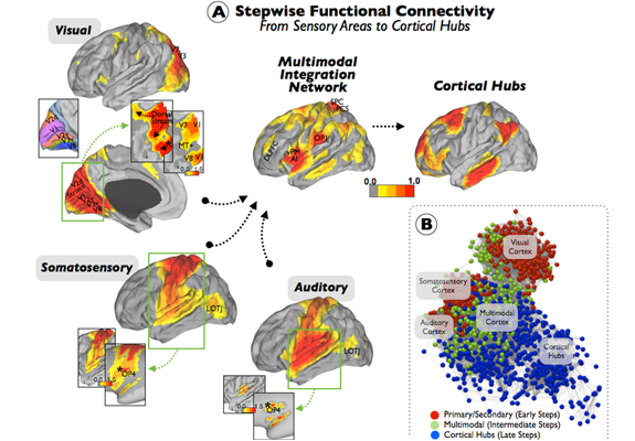
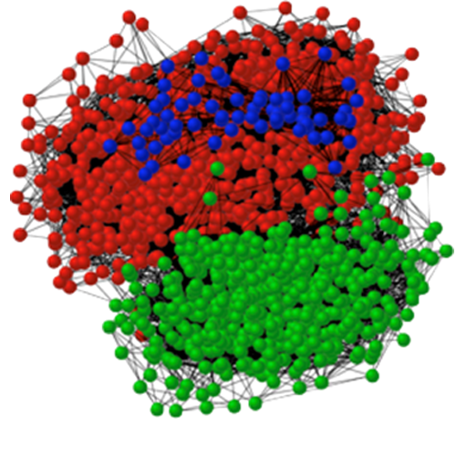
(A) Sensory functional streams of visual, somatosensory and auditory cortex revealed by Stepwise Functional Connectivity. Direct functional connectivity of visual cortex follows three different pathways, two dorsal and one ventromedial (inset, star). Two flat projections centered on the occipital lobe (green square) are presented using the same (top inset) and a relaxed color-scale threshold (bottom inset). Somatosensory cortex shows dense connections within the entire somatomotor cortex and strong direct connectivity with the secondary somatosensory area SII in OP4 (inset, star) and, to a lesser extent, to the LOTJ. Auditory cortex shows dense connections within the local auditory-related regions and strong direct connectivity with OP4 (inset, star) and, to a lesser extent, to the LOTJ. To better visualize the degree of connectivity on the target area (green square), conventional (bottom insets) and inflated projections (top insets) of the somatosensory and auditory results. In the subsequent steps of connectivity, all sensory modalities auditory reach a multimodal integration network and the cortical hubs of the human brain. Color scales are normalized z-scores of the stepwise degree of connectivity values. The visuotopic map (V1, V2, V3, V7, V8, and MT+) was provided by Caret software (Van Essen and Dierker, 2007). OP4: Operculum Parietale 4; OP1: Operculum Parietale 1; LOTJ: Lateral Occipito-Temporal Junction; DLPFC: Dorso-Lateral Prefrontal Cortex; SPC: Superior Parietal Cortex; PCS: Posterior central sulcus; vPM: Ventral Premotor; AI: Anterior Insula. (B) presents a network layout of the early (red nodes), intermediate (green nodes), and terminal (blue nodes) cores of the Stepwise Functional Connectivity analysis of the sensory cortex. The multimodal integration network (green nodes) acts as a network interface between the unimodal-related systems (red nodes) and the cortical hubs core (blue nodes).
An OP4 Functional Stream in the Language-Related Neuroarchitecture
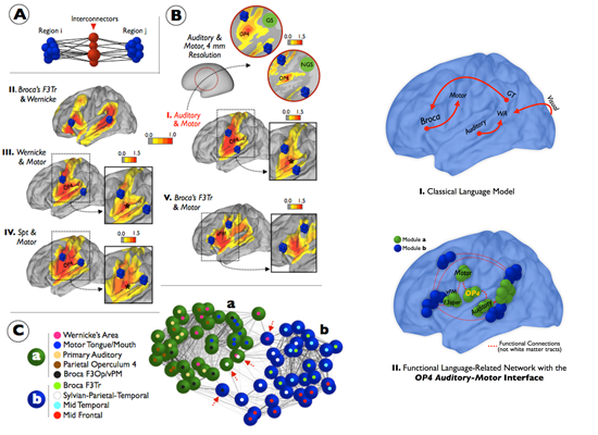
(A) shows a diagram of the network interconnectors method between region i and region j. (B) displays the results of the network interconnectors analysis between target regions. Star symbols (*) in magnified figures mark the maximum peak of the degree of interconnectivity value in OP4. Blue nodes in B represent the target regions under the study. Color scales are normalized z-scores of the stepwise degree of connectivity values. (C) shows the network layout of two modules (a: sensorimotor module in green; b: F3Tr-language-related module in blue) and overlapping regions between modules (namely, Spt, Wernicke’s area, and F3Op/vPM; red arrows) of the language-related network. Spt: Sylvian-Parietal-Temporal, F3Tr: pars triangularis of Broca’s complex; F3Op: pars opercularis of Broca’s complex; OP4: Operculum Parietale 4; vPM: Ventral Premotor.
Functional Streams and Cortical Integration in the Human Brain
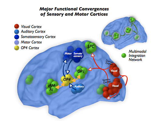
Diagram of the stepwise convergence of the sensory and motor systems into the multimodal integration network. OP4: Operculum Parietale 4; OP1: Operculum Parietale 1; SPC: Superior Parietal Cortex; vPM/aI: Ventral Premotor/Anterior Insula
In vivo Tau and Amyloid Network Profiles in the Aging Brain
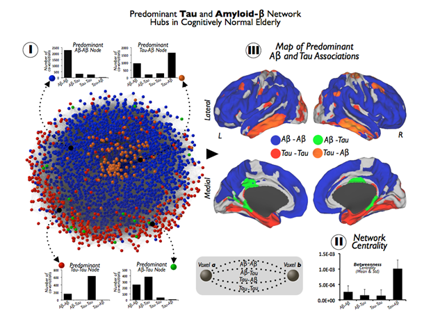
(I) shows the pathology network, based on all Tau and A interactions at the brain voxel-level. Network graph is displayed using a Kamada-Kawai energy layout in which the value of weighted links rank from 0 to 4, depending on the total number of significant associations for the pair of voxels (see diagram in middle-bottom for an schematic representation). The color partition of the network represents the predominant pathological associations in brain nodes. Column figures show random examples of connectivity profiles with a predominant association between Tau and A (Aβ-Aβ: blue nodes, Aβ-Tau: green nodes, Tau-Tau: red nodes, Tau-Aβ: orange nodes). (II) shows in a column figure the mean and SD of betweenness centrality in each predominant hub group of (I). (III) shows the projection of the brain nodes of the pathology network in (I) onto the cortical space.
Multimodal integration network in Blindness
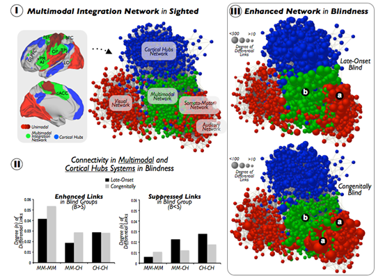
(A) Unimodal sensory cortices (different shades of red), multimodal integration cortices (green), and cortical hubs (blue) are displayed on template brains, as well as on a graph layout illustrating connectivity patterns across systems in the sighted controls. DLPFC = dorsolateral prefrontal cortex, FEF = frontal eye field, SPC = superior parietal cortex, vPM = ventral premotor, AI = anterior insula, OP = operculum parietale, TPJ = temporo-parietal junction, LO = lateral occipital, dACC = dorsal anterior cingulate cortex. (B) Bar graphs show statistically significant increased or enhanced (left) and decreased or suppressed (right) interconnectivity between multimodal regions and cortical hubs in blind subjects compared to MCs. Results in bar graphs show the total amount of FDR-corrected functional connections that differ between studied groups, and, thus, standard deviation or error bars do not apply here. MM = multimodal, CH = cortical hubs, B = Blind, S = Sighted. (C) Graph based layouts of the three hierarchical networks in LB and CB subjects [size of nodes = degree of differential links (DDL)]. Due to normalization and scale proportions, both segments of panel (C) were scaled to aid visualization. (a) unimodal cortices; (b) multimodal integration cortices