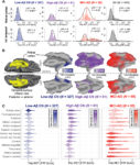
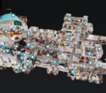
Some of the CMITT lab space is featured in a recently created virtual tour of the Bulfinch Building, celebrating 200 years of patient care at MGH. We are located in the basement, and if you click on the 6th highlight, you can start exploring from one of our laboratories
News Items
Dr Brugarolas receives ASN Jordi Folch-Pi Award
The ASN Jordi Folch-Pi Award is given to an outstanding young investigator demonstrating research competence and originality, significantly advancing our knowledge of neurochemistry, and showing a high degree of potential for future accomplishments. Dr. Brugarolas’ research focuses on developing novel PET ligands to understand the chemistry on the brain. He co-invented a tracer, [18F]3F4APk that binds to potassium channels in the brain, which are proteins that can signal demyelinated lesions.
SNMMI 2021 Young Investigator Awards
Three Gordon Center members, Felicitas Detmer, Gengyang Yuan, and Yanis Chemli, have received Young Investigator Award (YIA) recognition for their work presented at the Society of Nuclear Medicine and Molecular Imaging Annual Conference.
Dr. Hak Soo Choi Receives Distinguished Investigator Award from the Academy for Radiology & Biomedical Imaging Research
Hak Soo Choi, Ph.D., received the 2021 Distinguished Investigator Award from the Academy for Radiology & Biomedical Imaging Research.
Dr. Karla Ramos-Torres awarded PSDA grant funding
Dr. Ramos-Torres, a post-doctoral research fellow at the Gordon Center, was awarded an MGH Physician/Scientist Development award for “Imaging brain injury in animal models using PET.”
Conference abstract recognitions
Two Gordon Center students and a post-doctoral fellow have been recognized for their excellent work on recent conference abstracts.
Dr. Ruth Lim elected chair of the American Board of Nuclear Medicine
The cortical origin and initial spread of medial temporal tauopathy in Alzheimer’s disease assessed with positron emission tomography
Dr. Johnson’s Lab has reported on recent work, “The cortical origin and initial spread of medial temporal tauopathy in Alzheimer’s disease assessed with positron emission tomography”, using Positron emission tomography (PET) to image tau protein deposition in the brain in Alzheimer’s disease (AD).
Sanchez, J.S., et al. (2021) The cortical origin and initial spread of medial temporal tauopathy in Alzheimer’s disease assessed with positron emission tomography. Science Translational Medicine. doi.org/10.1126/scitranslmed.abc0655.