History
Inception of Positron Imaging at MGH
Before PET there was positron imaging. In 1950 Gordon Brownell suggested that coincidence detection could increase both the resolution and sensitivity of nuclear imaging of brain tumors. A simple positron scanner, using two opposed sodium iodide detectors, was quickly designed and built. The results were sufficiently encouraging that a more refined design followed in 1952.
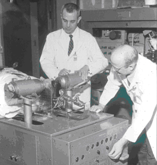
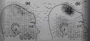
Figure 1 shows a picture of the scanner and Figure 2 shows the image formed by tapping with a solenoid on a piece of carbon paper with a rate proportional to the coincidence rate. By the early 1960’s more sensitive, multidetector designs were in use. More than 4000 brain scans were performed with coincidence detection and 74As between 1956 and 1964. The first area sensitive positron imaging device, PC-I was designed in 1968 and went into service in 1969-1970, used principally to study pulmonary physiology. In this same period, experiments were performed in several institutions investigating the feasibility of tomographic measurements and coincidence detection. David Chelser, a lab member working with Gordon Brownell, had already developed a reconstruction technique which we recognize today as the filtered back projection algorithm. Experiments with a turntable, and a pair of coincidence detectors which could be scanned in a rectilinear fashion showed that it was feasible to make quantitative image reconstructions of arbitrary activity distributions, the first practical 3D reconstruction. A second area sensitive camera, PC-II, was built to enable both rotation and translation of the detectors, that is specifically for the purpose of multi-slice three dimensional reconstruction.
Developments in kinetic measurements were carried on and supported by the rapid progress in instrumentation. Stimulated by Terry Jones, Subrumanium et al were among the first to investigate constant infusion of 15O-lableled compounds. Alpert reported the first multiple gated cardiac blood pool scans, a technique that gained considerable clinical importance in the 1970s-1990’s. David Chesler and his students investigated the noise power spectrum of CT, Pelc developed the first true 3D back projection algorithm. The noise properties of time-of-flight assisted reconstruction were investigated. Alpert et al, developed and demonstrated the first practical method for computing the local noise in positron emission tomography.
One of the most important contributions of the Physics Laboratory at the MGH was the idea of light-sharing to enable the use of very small crystals.
This was the enabling technology, existing to this day, in modern PET scanners. Prior to this development, a detector channel was a pair of sodium iodide crystals coupled to individual photo tubes. The size and geometry of the photo tubes limited the resolution and sensitivity of PET cameras and since increasing the number of crystals increased the number of photo tubes on a one-to-one basis high resolution PET was cost-prohibitive.
Charles A Burnham, an engineer in the Lab realized that an array of photo tubes could be used to find the center of brightness in a larger array of scintillation crystals. This principle was demonstrated in PR-I, a single ring detector with very high sensitivity and resolution. This principle quickly led to the commercial block detector which has enabled modern PET scanning.